 Movie
Movie Controller

[English] 日本語
 Yorodumi
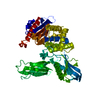
- PDB-3tg1: Crystal structure of p38alpha in complex with a MAPK docking partner -
+ Open data

- Basic information
| Entry | Database: PDB / ID: 3tg1 | ||||||
|---|---|---|---|---|---|---|---|
| Title | Crystal structure of p38alpha in complex with a MAPK docking partner | ||||||
 Components Components |
| ||||||
 Keywords Keywords | TRANSFERASE/HYDROLASE / Kinase/Rhodanese-like domain / docking interaction / TRANSFERASE-HYDROLASE complex | ||||||
| Function / homology |  Function and homology information Function and homology informationnegative regulation of epithelium regeneration / MAP kinase phosphatase activity / protein tyrosine/threonine phosphatase activity / MAP kinase tyrosine phosphatase activity / MAP kinase tyrosine/serine/threonine phosphatase activity / regulation of adaptive immune response / regulation of brown fat cell differentiation / negative regulation of epithelial cell migration / negative regulation of p38MAPK cascade / p38MAPK events ...negative regulation of epithelium regeneration / MAP kinase phosphatase activity / protein tyrosine/threonine phosphatase activity / MAP kinase tyrosine phosphatase activity / MAP kinase tyrosine/serine/threonine phosphatase activity / regulation of adaptive immune response / regulation of brown fat cell differentiation / negative regulation of epithelial cell migration / negative regulation of p38MAPK cascade / p38MAPK events / Activation of the AP-1 family of transcription factors / Platelet sensitization by LDL / RHO GTPases Activate NADPH Oxidases / ERK/MAPK targets / myoblast differentiation involved in skeletal muscle regeneration / Regulation of MITF-M-dependent genes involved in pigmentation / NLRP1 inflammasome complex assembly / activated TAK1 mediates p38 MAPK activation / ADP signalling through P2Y purinoceptor 1 / NOD1/2 Signaling Pathway / Oxidative Stress Induced Senescence / negative regulation of oligodendrocyte differentiation / Signaling by MAPK mutants / Regulation of TP53 Activity through Phosphorylation / Myogenesis / RAF-independent MAPK1/3 activation / JUN kinase binding / VEGFA-VEGFR2 Pathway / negative regulation of stress-activated MAPK cascade / dephosphorylation / negative regulation of JNK cascade / regulation of synaptic membrane adhesion / stress-induced premature senescence / positive regulation of regulatory T cell differentiation / stress-activated protein kinase signaling cascade / cell surface receptor protein serine/threonine kinase signaling pathway / positive regulation of myoblast fusion / cellular response to UV-B / mitogen-activated protein kinase p38 binding / cartilage condensation / NFAT protein binding / protein-serine/threonine phosphatase / positive regulation of myotube differentiation / regulation of cytokine production involved in inflammatory response / cellular response to lipoteichoic acid / p38MAPK cascade / response to dietary excess / response to muramyl dipeptide / fatty acid oxidation / oligodendrocyte differentiation / phosphatase activity / protein serine/threonine phosphatase activity / chondrocyte differentiation / MAP kinase activity / regulation of ossification / cellular response to vascular endothelial growth factor stimulus / mitogen-activated protein kinase / pyroptotic inflammatory response / vascular endothelial growth factor receptor signaling pathway / positive regulation of myoblast differentiation / negative regulation of hippo signaling / positive regulation of cardiac muscle cell proliferation / negative regulation of respiratory burst involved in inflammatory response / stress-activated MAPK cascade / skeletal muscle tissue development / positive regulation of interleukin-12 production / positive regulation of brown fat cell differentiation / striated muscle cell differentiation / response to muscle stretch / signal transduction in response to DNA damage / protein-tyrosine-phosphatase / Neutrophil degranulation / osteoclast differentiation / DNA damage checkpoint signaling / positive regulation of erythrocyte differentiation / lipopolysaccharide-mediated signaling pathway / placenta development / negative regulation of cell migration / positive regulation of D-glucose import across plasma membrane / tumor necrosis factor-mediated signaling pathway / cellular response to ionizing radiation / negative regulation of canonical Wnt signaling pathway / protein maturation / negative regulation of ERK1 and ERK2 cascade / positive regulation of protein import into nucleus / response to insulin / cellular response to virus / bone development / glucose metabolic process / negative regulation of epithelial cell proliferation / cell morphogenesis / cellular response to tumor necrosis factor / positive regulation of reactive oxygen species metabolic process / osteoblast differentiation / spindle pole / kinase activity / Negative regulation of MAPK pathway / MAPK cascade / cellular response to lipopolysaccharide / angiogenesis Similarity search - Function | ||||||
| Biological species |   Homo sapiens (human) Homo sapiens (human) | ||||||
| Method |  X-RAY DIFFRACTION / SYNCHROTRON / MOLECULAR REPLACEMENT / Resolution: 2.71 Å X-RAY DIFFRACTION / SYNCHROTRON / MOLECULAR REPLACEMENT / Resolution: 2.71 Å | ||||||
 Authors Authors | Zhang, Y.Y. / Wu, J.W. / Wang, Z.X. | ||||||
 Citation Citation | Journal: Sci.Signal. / Year: 2011 Title: A Distinct Interaction Mode Revealed by the Crystal Structure of the Kinase p38alpha with the MAPK Binding Domain of the Phosphatase MKP5. Authors: Zhang, Y.Y. / Wu, J.W. / Wang, Z.X. | ||||||
| History |
|
- Structure visualization
| Structure viewer | Molecule: MolmilJmol/JSmol |
|---|
- Downloads & links
-Download
| PDBx/mmCIF format | 3tg1.cif.gz | 205.2 KB | Display | PDBx/mmCIF format |
|---|---|---|---|---|
| PDB format | pdb3tg1.ent.gz | 164.4 KB | Display | PDB format |
| PDBx/mmJSON format | 3tg1.json.gz | Tree view | PDBx/mmJSON format | |
| Others |  Other downloads Other downloads |
-Validation report
| Arichive directory | https://data.pdbj.org/pub/pdb/validation_reports/tg/3tg1ftp://data.pdbj.org/pub/pdb/validation_reports/tg/3tg1 | HTTPS FTP |
|---|
-Related structure data
| Related structure data | 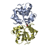 3tg3C  1p38  2oucS C: citing same article ( S: Starting model for refinement |
|---|---|
| Similar structure data |


-Links
 PDBj
PDBj


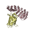
- Assembly
| Deposited unit | 
| ||||||||
|---|---|---|---|---|---|---|---|---|---|
| 1 |
| ||||||||
| Unit cell |
|
-Components
| #1: Protein | Mass: 43509.508 Da / Num. of mol.: 1 Source method: isolated from a genetically manipulated source Source: (gene. exp.)  References: UniProt: P47811, mitogen-activated protein kinase |
|---|---|
| #2: Protein | Mass: 18074.928 Da / Num. of mol.: 1 / Fragment: KBD (UNP RESIDUES 139-288) Source method: isolated from a genetically manipulated source Source: (gene. exp.) Homo sapiens (human) / Gene: DUSP10, MKP5 / Plasmid: pET-21b / Production host: References: UniProt: Q9Y6W6, protein-serine/threonine phosphatase, protein-tyrosine-phosphatase |
| #3: Water | ChemComp-HOH /  Mass: 18.015 Da / Num. of mol.: 15 / Source method: isolated from a natural source / Formula: H2O Mass: 18.015 Da / Num. of mol.: 15 / Source method: isolated from a natural source / Formula: H2O |
-Experimental details
-Experiment
| Experiment | Method: X-RAY DIFFRACTION / Number of used crystals: 1 |
|---|
- Sample preparation
| Crystal | Density Matthews: 2.41 Å3/Da / Density % sol: 48.92 % |
|---|---|
| Crystal grow | Temperature: 293 K / Method: vapor diffusion / pH: 7.5 Details: 100mM Tris pH7.5, 9% [w/v] polyethylene glycol 3350, 8% [w/v] sucrose, VAPOR DIFFUSION, temperature 293K |
-Data collection
| Diffraction | Mean temperature: 100 K |
|---|---|
| Diffraction source | Source: SYNCHROTRON / Site: SSRF  / Beamline: BL17U / Wavelength: 0.97924 Å / Beamline: BL17U / Wavelength: 0.97924 Å |
| Detector | Type: MARMOSAIC 225 mm CCD / Detector: CCD / Date: Jul 14, 2009 |
| Radiation | Monochromator: Si 111 / Protocol: SINGLE WAVELENGTH / Monochromatic (M) / Laue (L): M / Scattering type: x-ray |
| Radiation wavelength | Wavelength: 0.97924 Å / Relative weight: 1 |
| Reflection | Resolution: 2.7→36.214 Å / Num. obs: 17026 / % possible obs: 98.5 % / Observed criterion σ(F): 0 / Observed criterion σ(I): 0 / Redundancy: 6.8 % / Biso Wilson estimate: 71.3 Å2 / Rmerge(I) obs: 0.078 / Net I/σ(I): 20.6 |
| Reflection shell | Resolution: 2.7→2.8 Å / Redundancy: 5 % / Rmerge(I) obs: 0.359 / Mean I/σ(I) obs: 2.58 / Num. unique all: 1565 / % possible all: 93.3 |
- Processing
| Software |
| ||||||||||||||||||||||||||||||||||||||||||
|---|---|---|---|---|---|---|---|---|---|---|---|---|---|---|---|---|---|---|---|---|---|---|---|---|---|---|---|---|---|---|---|---|---|---|---|---|---|---|---|---|---|---|---|
| Refinement | Method to determine structure: MOLECULAR REPLACEMENT Starting model: 1P38, 2OUC Resolution: 2.71→36.214 Å / SU ML: 0.42 / Isotropic thermal model: Isotropic / Cross valid method: THROUGHOUT / σ(F): 1.34 / Phase error: 27.34 / Stereochemistry target values: ML
| ||||||||||||||||||||||||||||||||||||||||||
| Solvent computation | Shrinkage radii: 0.9 Å / VDW probe radii: 1.11 Å / Solvent model: FLAT BULK SOLVENT MODEL / Bsol: 69.609 Å2 / ksol: 0.337 e/Å3 | ||||||||||||||||||||||||||||||||||||||||||
| Displacement parameters | Biso mean: 88.8 Å2
| ||||||||||||||||||||||||||||||||||||||||||
| Refinement step | Cycle: LAST / Resolution: 2.71→36.214 Å
| ||||||||||||||||||||||||||||||||||||||||||
| Refine LS restraints |
| ||||||||||||||||||||||||||||||||||||||||||
| LS refinement shell | Refine-ID: X-RAY DIFFRACTION / Total num. of bins used: 6
|
