Method to determine structure: SAD / Resolution: 2.551→29.644 Å / SU ML: 0.4 / Cross valid method: THROUGHOUT / σ(F): 0.09 / Phase error: 28.62 / Stereochemistry target values: ML
Rfactor
Num. reflection
% reflection
Selection details
Rfree
0.2621
1156
10.17 %
RASOM
Rwork
0.1914
-
-
-
all
0.2017
11317
-
-
obs
0.1985
10161
81.3 %
-
Solvent computation
Shrinkage radii: 0.83 Å / VDW probe radii: 1.1 Å / Solvent model: FLAT BULK SOLVENT MODEL / Bsol: 81.258 Å2 / ksol: 0.354 e/Å3
Displacement parameters
Baniso -1
Baniso -2
Baniso -3
1-
-2.7951 Å2
-0 Å2
0 Å2
2-
-
-2.7951 Å2
-0 Å2
3-
-
-
5.5903 Å2
Refinement step
Cycle: LAST / Resolution: 2.551→29.644 Å
Protein
Nucleic acid
Ligand
Solvent
Total
Num. atoms
1896
0
0
32
1928
Refine LS restraints
Refine-ID
Type
Dev ideal
Number
X-RAY DIFFRACTION
f_bond_d
0.008
1914
X-RAY DIFFRACTION
f_angle_d
1.152
2562
X-RAY DIFFRACTION
f_dihedral_angle_d
20.267
746
X-RAY DIFFRACTION
f_chiral_restr
0.076
293
X-RAY DIFFRACTION
f_plane_restr
0.004
335
LS refinement shell
Resolution (Å)
Rfactor Rfree
Num. reflection Rfree
Rfactor Rwork
Num. reflection Rwork
Refine-ID
% reflection obs (%)
2.5508-2.6668
0.3416
129
0.2854
1176
X-RAY DIFFRACTION
75
2.6668-2.8073
0.3397
138
0.2509
1252
X-RAY DIFFRACTION
80
2.8073-2.983
0.2951
156
0.2268
1299
X-RAY DIFFRACTION
83
2.983-3.2131
0.3028
161
0.2072
1358
X-RAY DIFFRACTION
86
3.2131-3.536
0.2579
150
0.1724
1353
X-RAY DIFFRACTION
87
3.536-4.0466
0.2452
151
0.1632
1357
X-RAY DIFFRACTION
85
4.0466-5.094
0.2033
139
0.143
1293
X-RAY DIFFRACTION
83
5.094-29.6456
0.2876
132
0.24
1127
X-RAY DIFFRACTION
72
Refinement TLS params.
Method: refined / Origin x: -19.0184 Å / Origin y: -11.4072 Å / Origin z: 9.0294 Å
11
12
13
21
22
23
31
32
33
T
0.3396 Å2
0.0099 Å2
-0.0026 Å2
-
0.3009 Å2
-0.0087 Å2
-
-
0.4479 Å2
L
2.0548 °2
-0.0536 °2
-0.1311 °2
-
1.5149 °2
-0.1913 °2
-
-
0.1206 °2
S
-0.1851 Å °
-0.1543 Å °
0.1842 Å °
-0.2173 Å °
0.024 Å °
0.0459 Å °
0.0473 Å °
0.0235 Å °
0.1566 Å °
Refinement TLS group
Selection details: all
+
About Yorodumi
-
News
-
Feb 9, 2022. New format data for meta-information of EMDB entries
New format data for meta-information of EMDB entries
Version 3 of the EMDB header file is now the official format.
The previous official version 1.9 will be removed from the archive.
In the structure databanks used in Yorodumi, some data are registered as the other names, "COVID-19 virus" and "2019-nCoV". Here are the details of the virus and the list of structure data.
Jan 31, 2019. EMDB accession codes are about to change! (news from PDBe EMDB page)
EMDB accession codes are about to change! (news from PDBe EMDB page)
The allocation of 4 digits for EMDB accession codes will soon come to an end. Whilst these codes will remain in use, new EMDB accession codes will include an additional digit and will expand incrementally as the available range of codes is exhausted. The current 4-digit format prefixed with “EMD-” (i.e. EMD-XXXX) will advance to a 5-digit format (i.e. EMD-XXXXX), and so on. It is currently estimated that the 4-digit codes will be depleted around Spring 2019, at which point the 5-digit format will come into force.
The EM Navigator/Yorodumi systems omit the EMD- prefix.
Related info.:Q: What is EMD? / ID/Accession-code notation in Yorodumi/EM Navigator
Yorodumi is a browser for structure data from EMDB, PDB, SASBDB, etc.
This page is also the successor to EM Navigator detail page, and also detail information page/front-end page for Omokage search.
The word "yorodu" (or yorozu) is an old Japanese word meaning "ten thousand". "mi" (miru) is to see.
Related info.:EMDB / PDB / SASBDB / Comparison of 3 databanks / Yorodumi Search / Aug 31, 2016. New EM Navigator & Yorodumi / Yorodumi Papers / Jmol/JSmol / Function and homology information / Changes in new EM Navigator and Yorodumi
 Movie
Movie Controller

 Yorodumi
 Open data
 Basic information
 Components
 Keywords
 Function and homology information
 X-RAY DIFFRACTION /
  Authors
 Citation
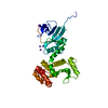
 Structure visualization
 Downloads & links
 Other downloads
 PDBj
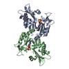
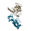
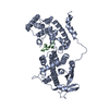
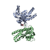
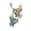
PDBj Assembly
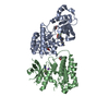
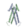

 Mass: 18.015 Da / Num. of mol.: 32 / Source method: isolated from a natural source / Formula: H2O
 Sample preparation
 Processing