Mass: 18.015 Da / Num. of mol.: 8 / Source method: isolated from a natural source / Formula: H2O
Sequence details
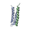
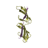
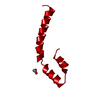
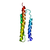
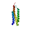
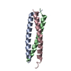
AUTHOR STATES THAT THE REGION CRYSTALLIZED IS ONLY PART OF ORIGINAL PROTEIN TARGET DUE TO IN-DROP ...AUTHOR STATES THAT THE REGION CRYSTALLIZED IS ONLY PART OF ORIGINAL PROTEIN TARGET DUE TO IN-DROP PROTEOLYSIS. THEY HAVE ATTEMPTED TO CRYSTALLIZE THE ENTIRE 137 AA PROTEIN, HOWEVER, MASS SPECTROSCOPIC ANALYSIS OF THE CRYSTALS REVEALED THAT THERE HAD BEEN SOME PROTEOLYSIS DURING THE CRYSTALLIZATION PROCESS, AND THE CRYSTAL ONLY HAD A MASS OF 4338 DA. THIS MASS CORRESPONDS TO THE COILED-COIL DOMAIN OF THE FULL LENGTH PROTEIN. SERRES RECORD IN THE PDB FILE REPRESETS UNP RESIDUES 699-737.
-
Experimental details
-
Experiment
Experiment
Method: X-RAY DIFFRACTION / Number of used crystals: 1
-
Sample preparation
Crystal
Density Matthews: 2.87 Å3/Da / Density % sol: 57.2 %
In the structure databanks used in Yorodumi, some data are registered as the other names, "COVID-19 virus" and "2019-nCoV". Here are the details of the virus and the list of structure data.
Jan 31, 2019. EMDB accession codes are about to change! (news from PDBe EMDB page)
EMDB accession codes are about to change! (news from PDBe EMDB page)
The allocation of 4 digits for EMDB accession codes will soon come to an end. Whilst these codes will remain in use, new EMDB accession codes will include an additional digit and will expand incrementally as the available range of codes is exhausted. The current 4-digit format prefixed with “EMD-” (i.e. EMD-XXXX) will advance to a 5-digit format (i.e. EMD-XXXXX), and so on. It is currently estimated that the 4-digit codes will be depleted around Spring 2019, at which point the 5-digit format will come into force.
The EM Navigator/Yorodumi systems omit the EMD- prefix.
Related info.:Q: What is EMD? / ID/Accession-code notation in Yorodumi/EM Navigator
Yorodumi is a browser for structure data from EMDB, PDB, SASBDB, etc.
This page is also the successor to EM Navigator detail page, and also detail information page/front-end page for Omokage search.
The word "yorodu" (or yorozu) is an old Japanese word meaning "ten thousand". "mi" (miru) is to see.
Related info.:EMDB / PDB / SASBDB / Comparison of 3 databanks / Yorodumi Search / Aug 31, 2016. New EM Navigator & Yorodumi / Yorodumi Papers / Jmol/JSmol / Function and homology information / Changes in new EM Navigator and Yorodumi
 Movie
Movie Controller

 Yorodumi
 Open data
 Basic information
 Components
 Keywords
 Function and homology information
 Homo sapiens (human)
 X-RAY DIFFRACTION /
  Authors
 Citation
 Structure visualization
 Downloads & links
 Other downloads
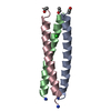


 PDBj
PDBj
 Assembly
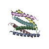


 Mass: 18.015 Da / Num. of mol.: 8 / Source method: isolated from a natural source / Formula: H2O
 Sample preparation
 / Beamline: 21-ID-G / Wavelength: 0.97857 Å
 Processing