 Movie
Movie Controller

[English] 日本語
 Yorodumi
- PDB-3t8k: The crystal structure of a functionally unknown protein Lebu_0176... -
+ Open data

- Basic information
| Entry | Database: PDB / ID: 3t8k | ||||||
|---|---|---|---|---|---|---|---|
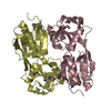
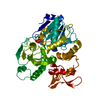
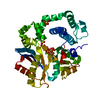
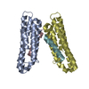
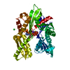
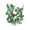
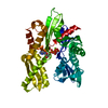
| Title | The crystal structure of a functionally unknown protein Lebu_0176 from Leptotrichia buccalis C-1013-b | ||||||
 Components Components | uncharacterized protein | ||||||
 Keywords Keywords | Structural Genomics / Unknown Function / PSI-Biology / protein structure initiative / midwest center f or structural genomics / MCSG / Midwest Center for Structural Genomics / alpha-hairpin repeat (Ankyrin repeat) fold | ||||||
| Function / homology | Ankyrin repeat-containing domain / Ankyrin repeat-containing domain superfamily / Serine Threonine Protein Phosphatase 5, Tetratricopeptide repeat / Alpha Horseshoe / Mainly Alpha / Uncharacterized protein Function and homology information Function and homology information | ||||||
| Biological species |  Leptotrichia buccalis (bacteria) Leptotrichia buccalis (bacteria) | ||||||
| Method |  X-RAY DIFFRACTION / SYNCHROTRON / SAD / Resolution: 1.769 Å X-RAY DIFFRACTION / SYNCHROTRON / SAD / Resolution: 1.769 Å | ||||||
 Authors Authors | Tan, K. / Bigelow, L. / Bearden, J. / Joachimiak, A. / Midwest Center for Structural Genomics (MCSG) | ||||||
 Citation Citation | Journal: To be Published Title: The crystal structure of a functionally unknown protein Lebu_0176 from Leptotrichia buccalis C-1013-b Authors: Tan, K. / Bigelow, L. / Bearden, J. / Joachimiak, A. | ||||||
| History |
|
- Structure visualization
| Structure viewer | Molecule: MolmilJmol/JSmol |
|---|
- Downloads & links
-Download
| PDBx/mmCIF format | 3t8k.cif.gz | 166.9 KB | Display | PDBx/mmCIF format |
|---|---|---|---|---|
| PDB format | pdb3t8k.ent.gz | 134.2 KB | Display | PDB format |
| PDBx/mmJSON format | 3t8k.json.gz | Tree view | PDBx/mmJSON format | |
| Others |  Other downloads Other downloads |
-Validation report
| Arichive directory | https://data.pdbj.org/pub/pdb/validation_reports/t8/3t8kftp://data.pdbj.org/pub/pdb/validation_reports/t8/3t8k | HTTPS FTP |
|---|
-Related structure data
| Similar structure data | |
|---|---|
| Other databases |


-Links
 PDBj
PDBj- Assembly
| Deposited unit | 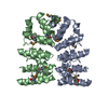
| ||||||||
|---|---|---|---|---|---|---|---|---|---|
| 1 |
| ||||||||
| Unit cell |
| ||||||||
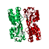
| Details | Experimentally unknown. It is predicted that the A and B chains form a dimer. |
-Components
| #1: Protein | Mass: 21771.219 Da / Num. of mol.: 2 Source method: isolated from a genetically manipulated source Source: (gene. exp.) Leptotrichia buccalis (bacteria) / Strain: C-1013-b / Gene: Lebu_0176 / Plasmid: pMCSG48 / Production host: #2: Water | ChemComp-HOH / |  Mass: 18.015 Da / Num. of mol.: 325 / Source method: isolated from a natural source / Formula: H2O Mass: 18.015 Da / Num. of mol.: 325 / Source method: isolated from a natural source / Formula: H2OHas protein modification | Y | |
|---|
-Experimental details
-Experiment
| Experiment | Method: X-RAY DIFFRACTION / Number of used crystals: 1 |
|---|
- Sample preparation
| Crystal | Density Matthews: 2.27 Å3/Da / Density % sol: 45.84 % |
|---|---|
| Crystal grow | Temperature: 277 K / Method: vapor diffusion, sitting drop / pH: 6.3 Details: 0.2M Ammonium Chloride, 20% (w/v) PEG 3350, pH 6.3, VAPOR DIFFUSION, SITTING DROP, temperature 277K |
-Data collection
| Diffraction | Mean temperature: 100 K |
|---|---|
| Diffraction source | Source: SYNCHROTRON / Site: APS  / Beamline: 19-ID / Wavelength: 0.97918 Å / Beamline: 19-ID / Wavelength: 0.97918 Å |
| Detector | Type: ADSC QUANTUM 315r / Detector: CCD / Date: Jul 31, 2011 / Details: Mirror |
| Radiation | Monochromator: Si 111 crystal / Protocol: SINGLE WAVELENGTH / Monochromatic (M) / Laue (L): M / Scattering type: x-ray |
| Radiation wavelength | Wavelength: 0.97918 Å / Relative weight: 1 |
| Reflection | Resolution: 1.769→34 Å / Num. all: 38999 / Num. obs: 38999 / % possible obs: 98.6 % / Observed criterion σ(F): 0 / Observed criterion σ(I): 0 / Redundancy: 6.7 % / Rmerge(I) obs: 0.104 / Net I/σ(I): 39.6 |
| Reflection shell | Resolution: 1.78→1.81 Å / Redundancy: 6.6 % / Mean I/σ(I) obs: 4.3 / Num. unique all: 1939 / % possible all: 98.7 |
- Processing
| Software |
| |||||||||||||||||||||||||||||||||||||||||||||||||||||||||||||||||||||||||||||||||||||||||||||||||||||||||
|---|---|---|---|---|---|---|---|---|---|---|---|---|---|---|---|---|---|---|---|---|---|---|---|---|---|---|---|---|---|---|---|---|---|---|---|---|---|---|---|---|---|---|---|---|---|---|---|---|---|---|---|---|---|---|---|---|---|---|---|---|---|---|---|---|---|---|---|---|---|---|---|---|---|---|---|---|---|---|---|---|---|---|---|---|---|---|---|---|---|---|---|---|---|---|---|---|---|---|---|---|---|---|---|---|---|---|
| Refinement | Method to determine structure: SAD / Resolution: 1.769→33.962 Å / SU ML: 0.38 / σ(F): 1.37 / Phase error: 21.6 / Stereochemistry target values: ML
| |||||||||||||||||||||||||||||||||||||||||||||||||||||||||||||||||||||||||||||||||||||||||||||||||||||||||
| Solvent computation | Shrinkage radii: 0.95 Å / VDW probe radii: 1.2 Å / Solvent model: FLAT BULK SOLVENT MODEL / Bsol: 30.463 Å2 / ksol: 0.321 e/Å3 | |||||||||||||||||||||||||||||||||||||||||||||||||||||||||||||||||||||||||||||||||||||||||||||||||||||||||
| Displacement parameters |
| |||||||||||||||||||||||||||||||||||||||||||||||||||||||||||||||||||||||||||||||||||||||||||||||||||||||||
| Refinement step | Cycle: LAST / Resolution: 1.769→33.962 Å
| |||||||||||||||||||||||||||||||||||||||||||||||||||||||||||||||||||||||||||||||||||||||||||||||||||||||||
| Refine LS restraints |
| |||||||||||||||||||||||||||||||||||||||||||||||||||||||||||||||||||||||||||||||||||||||||||||||||||||||||
| LS refinement shell |
| |||||||||||||||||||||||||||||||||||||||||||||||||||||||||||||||||||||||||||||||||||||||||||||||||||||||||
| Refinement TLS params. | Method: refined / Refine-ID: X-RAY DIFFRACTION
| |||||||||||||||||||||||||||||||||||||||||||||||||||||||||||||||||||||||||||||||||||||||||||||||||||||||||
| Refinement TLS group |
|
