 Movie
Movie Controller


+ Open data

- Basic information
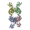
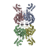
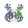
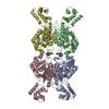
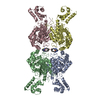
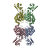
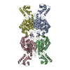
| Entry | Database: PDB / ID: 3ss5 | ||||||
|---|---|---|---|---|---|---|---|
| Title | Crystal structure of mouse Glutaminase C, L-glutamate-bound form | ||||||
 Components Components | Glutaminase C | ||||||
 Keywords Keywords | HYDROLASE / Glutaminase / L-Glutamine / Mitochondria / Catalysis product | ||||||
| Function / homology |  Function and homology information Function and homology informationGlutamate and glutamine metabolism / Glutamate Neurotransmitter Release Cycle / TP53 Regulates Metabolic Genes / L-glutamine catabolic process / intracellular glutamate homeostasis / regulation of respiratory gaseous exchange by nervous system process / glutaminase / : / glutaminase activity / suckling behavior ...Glutamate and glutamine metabolism / Glutamate Neurotransmitter Release Cycle / TP53 Regulates Metabolic Genes / L-glutamine catabolic process / intracellular glutamate homeostasis / regulation of respiratory gaseous exchange by nervous system process / glutaminase / : / glutaminase activity / suckling behavior / chemical synaptic transmission / protein homotetramerization / mitochondrial matrix / synapse / mitochondrion / identical protein binding / cytosol Similarity search - Function | ||||||
| Biological species |  | ||||||
| Method |  X-RAY DIFFRACTION / SYNCHROTRON / MOLECULAR REPLACEMENT / Resolution: 2.8 Å X-RAY DIFFRACTION / SYNCHROTRON / MOLECULAR REPLACEMENT / Resolution: 2.8 Å | ||||||
 Authors Authors | Ambrosio, A.L.B. / Dias, S.M.G. / Cerione, R.A. | ||||||
 Citation Citation | Journal: Proc.Natl.Acad.Sci.USA / Year: 2012 Title: Mitochondrial localization and structure-based phosphate activation mechanism of Glutaminase C with implications for cancer metabolism. Authors: Cassago, A. / Ferreira, A.P. / Ferreira, I.M. / Fornezari, C. / Gomes, E.R. / Greene, K.S. / Pereira, H.M. / Garratt, R.C. / Dias, S.M. / Ambrosio, A.L. | ||||||
| History |
|
- Structure visualization
| Structure viewer | Molecule: MolmilJmol/JSmol |
|---|
- Downloads & links
-Download
| PDBx/mmCIF format | 3ss5.cif.gz | 315.9 KB | Display | PDBx/mmCIF format |
|---|---|---|---|---|
| PDB format | pdb3ss5.ent.gz | 254.1 KB | Display | PDB format |
| PDBx/mmJSON format | 3ss5.json.gz | Tree view | PDBx/mmJSON format | |
| Others |  Other downloads Other downloads |
-Validation report
| Arichive directory | https://data.pdbj.org/pub/pdb/validation_reports/ss/3ss5ftp://data.pdbj.org/pub/pdb/validation_reports/ss/3ss5 | HTTPS FTP |
|---|
-Related structure data
| Related structure data |  3ss3C  3ss4C  3czdS C: citing same article ( S: Starting model for refinement |
|---|---|
| Similar structure data |


-Links
 PDBj
PDBj


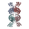
- Assembly
| Deposited unit | 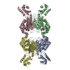
| ||||||||
|---|---|---|---|---|---|---|---|---|---|
| 1 |
| ||||||||
| Unit cell |
|
-Components
| #1: Protein | Mass: 53326.961 Da / Num. of mol.: 4 / Fragment: unp residues 134-609 Source method: isolated from a genetically manipulated source Source: (gene. exp.)  References: UniProt: Q69ZX9, UniProt: D3Z7P3*PLUS, glutaminase #2: Chemical | ChemComp-GLU /   Type: L-peptide linking / Mass: 147.129 Da / Num. of mol.: 4 / Source method: obtained synthetically / Formula: C5H9NO4 Type: L-peptide linking / Mass: 147.129 Da / Num. of mol.: 4 / Source method: obtained synthetically / Formula: C5H9NO4#3: Water | ChemComp-HOH / |  Mass: 18.015 Da / Num. of mol.: 214 / Source method: isolated from a natural source / Formula: H2O Mass: 18.015 Da / Num. of mol.: 214 / Source method: isolated from a natural source / Formula: H2O |
|---|
-Experimental details
-Experiment
| Experiment | Method: X-RAY DIFFRACTION / Number of used crystals: 1 |
|---|
- Sample preparation
| Crystal | Density Matthews: 2.93 Å3/Da / Density % sol: 58.03 % |
|---|---|
| Crystal grow | Temperature: 291 K / Method: vapor diffusion, sitting drop / pH: 6.5 Details: 17% PEG3350, 0.2M NaCl, 0.1M Bis-TRIS, pH 6.5, VAPOR DIFFUSION, SITTING DROP, temperature 291K |
-Data collection
| Diffraction | Mean temperature: 100 K |
|---|---|
| Diffraction source | Source: SYNCHROTRON / Site: LNLS  / Beamline: W01B-MX2 / Wavelength: 1.458 Å / Beamline: W01B-MX2 / Wavelength: 1.458 Å |
| Detector | Type: MARMOSAIC 225 mm CCD / Detector: CCD / Date: Sep 25, 2010 |
| Radiation | Monochromator: Si(111), Double Flat Crystal Monochromator with Fixed-exit Protocol: SINGLE WAVELENGTH / Monochromatic (M) / Laue (L): M / Scattering type: x-ray |
| Radiation wavelength | Wavelength: 1.458 Å / Relative weight: 1 |
| Reflection | Resolution: 2.8→42.88 Å / Num. all: 60426 / Num. obs: 59822 / % possible obs: 99 % / Observed criterion σ(F): 1.7 / Observed criterion σ(I): 1.7 / Redundancy: 2.3 % / Rmerge(I) obs: 0.106 / Rsym value: 0.106 / Net I/σ(I): 6 |
| Reflection shell | Resolution: 2.8→2.95 Å / Redundancy: 2.2 % / Mean I/σ(I) obs: 1.7 / Num. unique all: 8752 / Rsym value: 0.431 / % possible all: 98.7 |
- Processing
| Software |
| |||||||||||||||||||||||||||||||||||||||||||||||||||||||||||||||||||||||||||||||||||||||||||||||||||||||||||||||||||||||||||||||||||||||||||||||||||||||||||||||||
|---|---|---|---|---|---|---|---|---|---|---|---|---|---|---|---|---|---|---|---|---|---|---|---|---|---|---|---|---|---|---|---|---|---|---|---|---|---|---|---|---|---|---|---|---|---|---|---|---|---|---|---|---|---|---|---|---|---|---|---|---|---|---|---|---|---|---|---|---|---|---|---|---|---|---|---|---|---|---|---|---|---|---|---|---|---|---|---|---|---|---|---|---|---|---|---|---|---|---|---|---|---|---|---|---|---|---|---|---|---|---|---|---|---|---|---|---|---|---|---|---|---|---|---|---|---|---|---|---|---|---|---|---|---|---|---|---|---|---|---|---|---|---|---|---|---|---|---|---|---|---|---|---|---|---|---|---|---|---|---|---|---|---|
| Refinement | Method to determine structure: MOLECULAR REPLACEMENT Starting model: 3czd Resolution: 2.8→19.985 Å / SU ML: 0.82 / Cross valid method: THROUGHOUT / σ(F): 0 / Phase error: 26.39 / Stereochemistry target values: ML
| |||||||||||||||||||||||||||||||||||||||||||||||||||||||||||||||||||||||||||||||||||||||||||||||||||||||||||||||||||||||||||||||||||||||||||||||||||||||||||||||||
| Solvent computation | Shrinkage radii: 1.06 Å / VDW probe radii: 1.3 Å / Solvent model: FLAT BULK SOLVENT MODEL / Bsol: 23.793 Å2 / ksol: 0.316 e/Å3 | |||||||||||||||||||||||||||||||||||||||||||||||||||||||||||||||||||||||||||||||||||||||||||||||||||||||||||||||||||||||||||||||||||||||||||||||||||||||||||||||||
| Displacement parameters |
| |||||||||||||||||||||||||||||||||||||||||||||||||||||||||||||||||||||||||||||||||||||||||||||||||||||||||||||||||||||||||||||||||||||||||||||||||||||||||||||||||
| Refinement step | Cycle: LAST / Resolution: 2.8→19.985 Å
| |||||||||||||||||||||||||||||||||||||||||||||||||||||||||||||||||||||||||||||||||||||||||||||||||||||||||||||||||||||||||||||||||||||||||||||||||||||||||||||||||
| Refine LS restraints |
| |||||||||||||||||||||||||||||||||||||||||||||||||||||||||||||||||||||||||||||||||||||||||||||||||||||||||||||||||||||||||||||||||||||||||||||||||||||||||||||||||
| LS refinement shell |
|
