Mass: 18.015 Da / Num. of mol.: 193 / Source method: isolated from a natural source / Formula: H2O
Sequence details
1. THE CONSTRUCT WAS EXPRESSED WITH AN N-TERMINAL PURIFICATION TAG MGSDKIHHHHHHENLYFQG. THE TAG WAS ...1. THE CONSTRUCT WAS EXPRESSED WITH AN N-TERMINAL PURIFICATION TAG MGSDKIHHHHHHENLYFQG. THE TAG WAS REMOVED WITH TEV PROTEASE LEAVING ONLY A GLYCINE (0) FOLLOWED BY RESIDUES 490-600 OF THE TARGET SEQUENCE. 2. THE PROTEIN WAS REDUCTIVELY METHYLATED PRIOR TO CRYSTALLIZATION.
-
Experimental details
-
Experiment
Experiment
Method: X-RAY DIFFRACTION / Number of used crystals: 1
-
Sample preparation
Crystal
Density Matthews: 2.26 Å3/Da / Density % sol: 45.47 %
Resolution: 1.7→28.351 Å / Num. all: 26440 / Num. obs: 26440 / % possible obs: 100 % / Redundancy: 7.1 % / Rsym value: 0.067 / Net I/σ(I): 15.8
Reflection shell
Diffraction-ID: 1
Resolution (Å)
Redundancy (%)
Rmerge(I) obs
Mean I/σ(I) obs
Num. measured all
Num. unique all
Rsym value
% possible all
1.7-1.74
7.1
0.636
1.2
13539
1904
0.636
100
1.74-1.79
7.1
0.533
1.5
13337
1866
0.533
100
1.79-1.84
7.1
0.434
1.8
12957
1813
0.434
100
1.84-1.9
7.2
0.323
2.4
12639
1762
0.323
100
1.9-1.96
7.2
0.24
3.2
12313
1716
0.24
100
1.96-2.03
7.2
0.194
4
12026
1674
0.194
100
2.03-2.11
7.2
0.155
5
11475
1596
0.155
100
2.11-2.19
7.2
0.132
5.6
11280
1571
0.132
100
2.19-2.29
7.2
0.126
5.7
10527
1466
0.126
100
2.29-2.4
7.2
0.121
5.9
10248
1426
0.121
100
2.4-2.53
7.2
0.113
6.3
9843
1373
0.113
100
2.53-2.69
7.2
0.095
7.3
9277
1294
0.095
100
2.69-2.87
7.1
0.077
8.9
8709
1220
0.077
100
2.87-3.1
7.1
0.064
10.2
8150
1149
0.064
100
3.1-3.4
7.1
0.05
13.1
7534
1065
0.05
100
3.4-3.8
7
0.038
17.1
6766
969
0.038
100
3.8-4.39
6.9
0.039
16.3
5981
863
0.039
100
4.39-5.38
6.8
0.034
18.3
5080
750
0.034
100
5.38-7.6
6.5
0.041
15.9
3925
603
0.041
100
7.6-28.351
5.7
0.036
17.2
2046
360
0.036
97.9
-
Phasing
Phasing
Method: MAD
-
Processing
Software
Name
Version
Classification
NB
MolProbity
3beta29
modelbuilding
PDB_EXTRACT
3.1
dataextraction
SHELX
phasing
SHARP
phasing
SCALA
3.3.15
datascaling
BUSTER-TNT
2.8.0
refinement
MOSFLM
datareduction
SHELXD
phasing
BUSTER
2.8.0
refinement
Refinement
Method to determine structure: MAD / Resolution: 1.7→28.351 Å / Cor.coef. Fo:Fc: 0.9347 / Cor.coef. Fo:Fc free: 0.9261 / Occupancy max: 1 / Occupancy min: 0.25 / Cross valid method: THROUGHOUT / σ(F): 0 Details: 1. A MET-INHIBITION PROTOCOL WAS USED FOR SELENOMETHIONINE INCORPORATION DURING PROTEIN EXPRESSION. THE OCCUPANCY OF THE SE ATOMS IN THE MSE RESIDUES WAS REDUCED TO 0.75 FOR THE REDUCED ...Details: 1. A MET-INHIBITION PROTOCOL WAS USED FOR SELENOMETHIONINE INCORPORATION DURING PROTEIN EXPRESSION. THE OCCUPANCY OF THE SE ATOMS IN THE MSE RESIDUES WAS REDUCED TO 0.75 FOR THE REDUCED SCATTERING POWER DUE TO PARTIAL S-MET INCORPORATION. 2. ATOM RECORD CONTAINS SUM OF TLS AND RESIDUAL B FACTORS. ANISOU RECORD CONTAINS SUM OF TLS AND RESIDUAL U FACTORS. 3. SULFATE (SO4) AND CHLORIDE (CL) FROM THE CRYSTALLIZATION AND THE PURIFICATION BUFFER RESPECTIVELY HAVE BEEN MODELED INTO THE STRUCTURE. 4. NCS RESTRAINTS WERE APPLIED USING BUSTER'S LSSR RESTRAINT REPRESENTATION (-AUTONCS). 5. THE REFINEMENT WAS RESTRAINED AGAINST THE MAD PHASES. 6.THE PROTEIN WAS SUBJECTED TO REDUCTIVE METHYLATION PRIOR TO CRYSTALLIZATION AND LYSINES HAVE BEEN MODELED AS N-DIMETHYL-LYSINE (MLY). 7. ELECTON DENSITY INDICATES THAT THE N-TERMINAL GLYCINE RESIDUE (GLY 0) ON SUBUNIT B IS DI-METHYLATED; THEREFORE, THIS RESIDUE WAS MODELED AS DIMETHYL GLYCINE (DMG). 8. DIFFERENCE ELECTRON DENSITY IN A CRYSTAL PACKING INTERFACE NEAR GLU 547 COULD NOT BE RELIABLY ASSIGNED AND WAS NOT MODELED.
In the structure databanks used in Yorodumi, some data are registered as the other names, "COVID-19 virus" and "2019-nCoV". Here are the details of the virus and the list of structure data.
Jan 31, 2019. EMDB accession codes are about to change! (news from PDBe EMDB page)
EMDB accession codes are about to change! (news from PDBe EMDB page)
The allocation of 4 digits for EMDB accession codes will soon come to an end. Whilst these codes will remain in use, new EMDB accession codes will include an additional digit and will expand incrementally as the available range of codes is exhausted. The current 4-digit format prefixed with “EMD-” (i.e. EMD-XXXX) will advance to a 5-digit format (i.e. EMD-XXXXX), and so on. It is currently estimated that the 4-digit codes will be depleted around Spring 2019, at which point the 5-digit format will come into force.
The EM Navigator/Yorodumi systems omit the EMD- prefix.
Related info.:Q: What is EMD? / ID/Accession-code notation in Yorodumi/EM Navigator
Yorodumi is a browser for structure data from EMDB, PDB, SASBDB, etc.
This page is also the successor to EM Navigator detail page, and also detail information page/front-end page for Omokage search.
The word "yorodu" (or yorozu) is an old Japanese word meaning "ten thousand". "mi" (miru) is to see.
Related info.:EMDB / PDB / SASBDB / Comparison of 3 databanks / Yorodumi Search / Aug 31, 2016. New EM Navigator & Yorodumi / Yorodumi Papers / Jmol/JSmol / Function and homology information / Changes in new EM Navigator and Yorodumi
 Movie
Movie Controller

 Yorodumi
 Open data
 Basic information
 Components
 Keywords
 Function and homology information
 X-RAY DIFFRACTION /
  Authors
 Citation
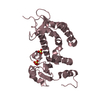
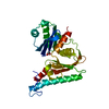
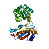
 Structure visualization
 Downloads & links
 Other downloads


 PDBj
PDBj


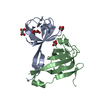
 Assembly


 Mass: 96.063 Da / Num. of mol.: 2 / Source method: obtained synthetically / Formula: SO4
 Mass: 35.453 Da / Num. of mol.: 4 / Source method: obtained synthetically / Formula: Cl
 Mass: 18.015 Da / Num. of mol.: 193 / Source method: isolated from a natural source / Formula: H2O
 Sample preparation
 / Beamline: BL9-2 / Wavelength: 0.91837,0.9792,0.97892
 Processing