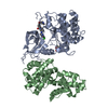
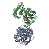
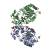
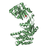
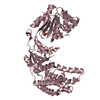
CRYSTAL PACKING ANALYSIS AND SIZE-EXCLUSION CHROMATOGRAPHY IN COMBINATION WITH STATIC LIGHT SCATTERING SUPPORT THE ASSIGNMENT OF A DIMER AS THE SIGNIFICANT OLIGOMERIZATION STATE IN SOLUTION.
Mass: 18.015 Da / Num. of mol.: 187 / Source method: isolated from a natural source / Formula: H2O
Has protein modification
Y
Sequence details
THE CONSTRUCT WAS EXPRESSED WITH A PURIFICATION TAG MGSDKIHHHHHHENLYFQG. THE TAG WAS REMOVED WITH ...THE CONSTRUCT WAS EXPRESSED WITH A PURIFICATION TAG MGSDKIHHHHHHENLYFQG. THE TAG WAS REMOVED WITH TEV PROTEASE LEAVING ONLY A GLYCINE (0) FOLLOWED BY THE TARGET SEQUENCE.
-
Experimental details
-
Experiment
Experiment
Method: X-RAY DIFFRACTION / Number of used crystals: 1
-
Sample preparation
Crystal
Density Matthews: 1.98 Å3/Da / Density % sol: 37.93 % Description: DATA WERE SCALED USING XSCALE WITH FRIEDEL PAIRS KEPT AS SEPARATE WHEN COMPUTING R MERGE, COMPLETENESS AND
Crystal grow
Temperature: 277 K / Method: vapor diffusion, sitting drop / pH: 3.64 Details: 10.0% 2-methyl-2,4-pentanediol, 0.1M citric acid pH 3.64, NANODROP, VAPOR DIFFUSION, SITTING DROP, temperature 277K
Resolution: 1.6→28.757 Å / Num. obs: 34491 / % possible obs: 92.4 % / Observed criterion σ(I): -3 / Biso Wilson estimate: 26.711 Å2 / Rmerge(I) obs: 0.027 / Net I/σ(I): 12.53
Reflection shell
Diffraction-ID: 1
Resolution (Å)
Highest resolution (Å)
Rmerge(I) obs
Mean I/σ(I) obs
Num. measured obs
Num. unique obs
% possible all
1.6-1.66
0.456
1.7
7588
5736
79.5
1.66-1.72
0.377
2
7739
5743
91.5
1.72-1.8
0.233
3
8879
6601
92.2
1.8-1.9
0.145
4.6
9136
6797
93.3
1.9-2.02
0.076
7.7
8778
6568
93.5
2.02-2.17
0.05
11.2
8305
6240
94.4
2.17-2.39
0.035
15
8870
6684
95.3
2.39-2.73
0.027
19.7
8591
6497
95.2
2.73-3.44
0.019
26.4
8912
6743
96.2
3.44
0.018
31.3
8533
6515
93
-
Phasing
Phasing
Method: MAD
-
Processing
Software
Name
Version
Classification
NB
MolProbity
3beta29
modelbuilding
PDB_EXTRACT
3.1
dataextraction
SHELX
phasing
SHARP
phasing
XSCALE
January30, 2009
datascaling
BUSTER-TNT
2.8.0
refinement
XDS
datareduction
SHELXD
phasing
BUSTER
2.8.0
refinement
Refinement
Method to determine structure: MAD / Resolution: 1.6→28.757 Å / Cor.coef. Fo:Fc: 0.93 / Cor.coef. Fo:Fc free: 0.9227 / Occupancy max: 1 / Occupancy min: 0.37 / Cross valid method: THROUGHOUT / σ(F): 0 Details: 1. A MET-INHIBITION PROTOCOL WAS USED FOR SELENOMETHIONINE INCORPORATION DURING PROTEIN EXPRESSION. THE OCCUPANCY OF THE SE ATOMS IN THE MSE RESIDUES WAS REDUCED TO 0.75 FOR THE REDUCED ...Details: 1. A MET-INHIBITION PROTOCOL WAS USED FOR SELENOMETHIONINE INCORPORATION DURING PROTEIN EXPRESSION. THE OCCUPANCY OF THE SE ATOMS IN THE MSE RESIDUES WAS REDUCED TO 0.75 FOR THE REDUCED SCATTERING POWER DUE TO PARTIAL S-MET INCORPORATION. 2. ATOM RECORD CONTAINS SUM OF TLS AND RESIDUAL B FACTORS. ANISOU RECORD CONTAINS SUM OF TLS AND RESIDUAL U FACTORS. 3. 1,2 ETHANEDIOL (EDO) FROM THE CRYOPROTECTANT HAS BEEN MODELED IN THE SOLVENT STRUCTURE. 4. RAMACHANDRAN OUTLIER AT RESIDUE 294 IS IN A REGION OF SUB-OPTIMAL ELECTRON DENSITY. 5. THE STRUCTURE HAS SEVERAL UNMODELED REGIONS WHICH HAVE ELECTRON DENSITY THAT COULD NOT BE RELIABLY INTERPRETED. IN ADDITION, THERE ARE THREE REGIONS OF SUB-OPTIMAL MODEL FIT IN WHICH THE MOST STRUCTURALLY SIMILAR PROTEIN (AS DETERMINED BY THE PROGRAM SSM), PDB ID 5DAA, WAS USED TO GUIDE THE MODELING. THESE INCLUDE RESIDUES 143-150, 204-221 AND 291-295. 6. THE REFINEMENT WAS RESTRAINED WITH THE MAD PHASES.
In the structure databanks used in Yorodumi, some data are registered as the other names, "COVID-19 virus" and "2019-nCoV". Here are the details of the virus and the list of structure data.
Jan 31, 2019. EMDB accession codes are about to change! (news from PDBe EMDB page)
EMDB accession codes are about to change! (news from PDBe EMDB page)
The allocation of 4 digits for EMDB accession codes will soon come to an end. Whilst these codes will remain in use, new EMDB accession codes will include an additional digit and will expand incrementally as the available range of codes is exhausted. The current 4-digit format prefixed with “EMD-” (i.e. EMD-XXXX) will advance to a 5-digit format (i.e. EMD-XXXXX), and so on. It is currently estimated that the 4-digit codes will be depleted around Spring 2019, at which point the 5-digit format will come into force.
The EM Navigator/Yorodumi systems omit the EMD- prefix.
Related info.:Q: What is EMD? / ID/Accession-code notation in Yorodumi/EM Navigator
Yorodumi is a browser for structure data from EMDB, PDB, SASBDB, etc.
This page is also the successor to EM Navigator detail page, and also detail information page/front-end page for Omokage search.
The word "yorodu" (or yorozu) is an old Japanese word meaning "ten thousand". "mi" (miru) is to see.
Related info.:EMDB / PDB / SASBDB / Comparison of 3 databanks / Yorodumi Search / Aug 31, 2016. New EM Navigator & Yorodumi / Yorodumi Papers / Jmol/JSmol / Function and homology information / Changes in new EM Navigator and Yorodumi
 Movie
Movie Controller

 Yorodumi
 Open data
 Basic information
 Components
 Keywords
 Function and homology information
 Corynebacterium glutamicum (bacteria)
 X-RAY DIFFRACTION /
  Authors
 Citation
 Structure visualization
 Downloads & links
 Other downloads
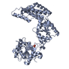


 PDBj
PDBj
 Assembly


 Mass: 62.068 Da / Num. of mol.: 3 / Source method: obtained synthetically / Formula: C2H6O2
 Mass: 18.015 Da / Num. of mol.: 187 / Source method: isolated from a natural source / Formula: H2O
 Sample preparation
 / Beamline: BL9-2 / Wavelength: 0.91837,0.97949,0.97932
 Processing