 Movie
Movie Controller

[English] 日本語
 Yorodumi
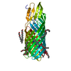
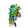
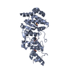
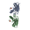
- PDB-3slj: Pre-cleavage Structure of the Autotransporter EspP - N1023A mutant -
+ Open data

- Basic information
| Entry | Database: PDB / ID: 3slj | ||||||
|---|---|---|---|---|---|---|---|
| Title | Pre-cleavage Structure of the Autotransporter EspP - N1023A mutant | ||||||
 Components Components | Serine protease espP | ||||||
 Keywords Keywords | PROTEIN TRANSPORT / beta barrel / membrane protein / asparagine cyclization / autocleavage | ||||||
| Function / homology |  Function and homology information Function and homology informationtranslocation of peptides or proteins into host / Secretion of toxins / Hydrolases; Acting on peptide bonds (peptidases); Serine endopeptidases / cell outer membrane / periplasmic space / serine-type endopeptidase activity / cell surface / proteolysis / extracellular region Similarity search - Function | ||||||
| Biological species |  | ||||||
| Method |  X-RAY DIFFRACTION / SYNCHROTRON / MOLECULAR REPLACEMENT / molecular replacement / Resolution: 2.481 Å X-RAY DIFFRACTION / SYNCHROTRON / MOLECULAR REPLACEMENT / molecular replacement / Resolution: 2.481 Å | ||||||
 Authors Authors | Barnard, T.B. / Noinaj, N. / Easley, N.C. / Kuszak, A.J. / Buchanan, S.K. | ||||||
 Citation Citation | Journal: J.Mol.Biol. / Year: 2012 Title: Molecular basis for the activation of a catalytic asparagine residue in a self-cleaving bacterial autotransporter. Authors: Barnard, T.J. / Gumbart, J. / Peterson, J.H. / Noinaj, N. / Easley, N.C. / Dautin, N. / Kuszak, A.J. / Tajkhorshid, E. / Bernstein, H.D. / Buchanan, S.K. | ||||||
| History |
|
- Structure visualization
| Structure viewer | Molecule: MolmilJmol/JSmol |
|---|
- Downloads & links
-Download
| PDBx/mmCIF format | 3slj.cif.gz | 135.5 KB | Display | PDBx/mmCIF format |
|---|---|---|---|---|
| PDB format | pdb3slj.ent.gz | 104.4 KB | Display | PDB format |
| PDBx/mmJSON format | 3slj.json.gz | Tree view | PDBx/mmJSON format | |
| Others |  Other downloads Other downloads |
-Validation report
| Arichive directory | https://data.pdbj.org/pub/pdb/validation_reports/sl/3sljftp://data.pdbj.org/pub/pdb/validation_reports/sl/3slj | HTTPS FTP |
|---|
-Related structure data
| Related structure data |  3sloC  3sltC  2qomS C: citing same article ( S: Starting model for refinement |
|---|---|
| Similar structure data |


-Links
 PDBj
PDBj- Assembly
| Deposited unit | 
| ||||||||
|---|---|---|---|---|---|---|---|---|---|
| 1 |
| ||||||||
| Unit cell |
|
-Components
| #1: Protein | Mass: 34417.996 Da / Num. of mol.: 1 Fragment: Autotransporter protein espP translocator (UNP residues 999-1300) Mutation: N1023A Source method: isolated from a genetically manipulated source Source: (gene. exp.) | ||
|---|---|---|---|
| #2: Chemical | ChemComp-C8E / (   Mass: 306.438 Da / Num. of mol.: 9 / Source method: obtained synthetically / Formula: C16H34O5 / Comment: C8E, detergent*YM Mass: 306.438 Da / Num. of mol.: 9 / Source method: obtained synthetically / Formula: C16H34O5 / Comment: C8E, detergent*YM#3: Water | ChemComp-HOH / |  Mass: 18.015 Da / Num. of mol.: 55 / Source method: isolated from a natural source / Formula: H2O Mass: 18.015 Da / Num. of mol.: 55 / Source method: isolated from a natural source / Formula: H2O |
-Experimental details
-Experiment
| Experiment | Method: X-RAY DIFFRACTION / Number of used crystals: 1 |
|---|
- Sample preparation
| Crystal | Density Matthews: 3.41 Å3/Da / Density % sol: 63.96 % |
|---|---|
| Crystal grow | Temperature: 294 K / pH: 7.5 Details: 20% w/v PEG8000, 20% v/v glycerol, pH 7.5, HANGING DROP, temperature 294K |
-Data collection
| Diffraction | Mean temperature: 100 K | |||||||||||||||||||||||||||||||||||||||||||||||||||||||||||||||||||||||||||||
|---|---|---|---|---|---|---|---|---|---|---|---|---|---|---|---|---|---|---|---|---|---|---|---|---|---|---|---|---|---|---|---|---|---|---|---|---|---|---|---|---|---|---|---|---|---|---|---|---|---|---|---|---|---|---|---|---|---|---|---|---|---|---|---|---|---|---|---|---|---|---|---|---|---|---|---|---|---|---|
| Diffraction source | Source: SYNCHROTRON / Site: APS  / Beamline: 22-ID / Wavelength: 1 / Beamline: 22-ID / Wavelength: 1 | |||||||||||||||||||||||||||||||||||||||||||||||||||||||||||||||||||||||||||||
| Detector | Type: MARMOSAIC 300 mm CCD / Detector: CCD / Date: Apr 18, 2009 / Details: mirrors | |||||||||||||||||||||||||||||||||||||||||||||||||||||||||||||||||||||||||||||
| Radiation | Monochromator: Si 220 (ROSENBAUM-ROCK DOUBLE-CRYSTAL MONOCHROMATOR) Protocol: SINGLE WAVELENGTH / Monochromatic (M) / Laue (L): M / Scattering type: x-ray | |||||||||||||||||||||||||||||||||||||||||||||||||||||||||||||||||||||||||||||
| Radiation wavelength | Wavelength: 1 Å / Relative weight: 1 | |||||||||||||||||||||||||||||||||||||||||||||||||||||||||||||||||||||||||||||
| Reflection | Resolution: 2.48→50 Å / Num. all: 17548 / Num. obs: 17443 / % possible obs: 99.4 % / Observed criterion σ(I): -3 / Redundancy: 4.8 % / Rmerge(I) obs: 0.087 / Χ2: 1.04 / Net I/σ(I): 9.5 | |||||||||||||||||||||||||||||||||||||||||||||||||||||||||||||||||||||||||||||
| Reflection shell |
|
-Phasing
| Phasing | Method: molecular replacement | |||||||||
|---|---|---|---|---|---|---|---|---|---|---|
| Phasing MR | Rfactor: 50.9 / Model details: Phaser MODE: MR_AUTO
|
- Processing
| Software |
| ||||||||||||||||||||||||||||||||||||||||||||||||||||||||||||||||||||||||
|---|---|---|---|---|---|---|---|---|---|---|---|---|---|---|---|---|---|---|---|---|---|---|---|---|---|---|---|---|---|---|---|---|---|---|---|---|---|---|---|---|---|---|---|---|---|---|---|---|---|---|---|---|---|---|---|---|---|---|---|---|---|---|---|---|---|---|---|---|---|---|---|---|---|
| Refinement | Method to determine structure: MOLECULAR REPLACEMENT Starting model: PDB ENTRY 2QOM Resolution: 2.481→43.426 Å / Occupancy max: 1 / Occupancy min: 0.41 / FOM work R set: 0.8267 / SU ML: 0.35 / σ(F): 1.34 / Phase error: 23.13 / Stereochemistry target values: ML
| ||||||||||||||||||||||||||||||||||||||||||||||||||||||||||||||||||||||||
| Solvent computation | Shrinkage radii: 0.9 Å / VDW probe radii: 1.11 Å / Solvent model: FLAT BULK SOLVENT MODEL / Bsol: 63.558 Å2 / ksol: 0.36 e/Å3 | ||||||||||||||||||||||||||||||||||||||||||||||||||||||||||||||||||||||||
| Displacement parameters | Biso max: 99.98 Å2 / Biso mean: 46.1032 Å2 / Biso min: 18.96 Å2
| ||||||||||||||||||||||||||||||||||||||||||||||||||||||||||||||||||||||||
| Refinement step | Cycle: LAST / Resolution: 2.481→43.426 Å
| ||||||||||||||||||||||||||||||||||||||||||||||||||||||||||||||||||||||||
| Refine LS restraints |
| ||||||||||||||||||||||||||||||||||||||||||||||||||||||||||||||||||||||||
| LS refinement shell | Refine-ID: X-RAY DIFFRACTION / Total num. of bins used: 6
| ||||||||||||||||||||||||||||||||||||||||||||||||||||||||||||||||||||||||
| Refinement TLS params. | S33: 0 Å ° / Method: refined / Refine-ID: X-RAY DIFFRACTION
| ||||||||||||||||||||||||||||||||||||||||||||||||||||||||||||||||||||||||
| Refinement TLS group |
|
