 Movie
Movie Controller

[English] 日本語
 Yorodumi
- PDB-3sk2: Crystal structure of phenazine resistance protein EhpR from Enter... -
+ Open data

- Basic information
| Entry | Database: PDB / ID: 3sk2 | ||||||
|---|---|---|---|---|---|---|---|
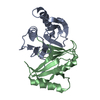
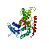
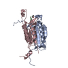
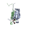
| Title | Crystal structure of phenazine resistance protein EhpR from Enterobacter agglomerans (Erwinia herbicola, Pantoea agglomerans) Eh1087 in complex with griseoluteic acid | ||||||
 Components Components | EhpR | ||||||
 Keywords Keywords | GRISEOLUTEATE-BINDING PROTEIN / antibiotic resistance | ||||||
| Function / homology |  Function and homology information Function and homology informationorganic acid binding / response to antibiotic / protein homodimerization activity Similarity search - Function | ||||||
| Biological species |  Pantoea agglomerans (bacteria) Pantoea agglomerans (bacteria) | ||||||
| Method |  X-RAY DIFFRACTION / SYNCHROTRON / MOLECULAR REPLACEMENT / Resolution: 1.01 Å X-RAY DIFFRACTION / SYNCHROTRON / MOLECULAR REPLACEMENT / Resolution: 1.01 Å | ||||||
 Authors Authors | Blankenfeldt, W. / Yu, S. | ||||||
 Citation Citation | Journal: Bmc Struct.Biol. / Year: 2011 Title: Atomic resolution structure of EhpR: phenazine resistance in Enterobacter agglomerans Eh1087 follows principles of bleomycin / mitomycin C resistance in other bacteria. Authors: Yu, S. / Vit, A. / Devenish, S. / Mahanty, H.K. / Itzen, A. / Goody, R.S. / Blankenfeldt, W. | ||||||
| History |
|
- Structure visualization
| Structure viewer | Molecule: MolmilJmol/JSmol |
|---|
- Downloads & links
-Download
| PDBx/mmCIF format | 3sk2.cif.gz | 151.1 KB | Display | PDBx/mmCIF format |
|---|---|---|---|---|
| PDB format | pdb3sk2.ent.gz | 120.8 KB | Display | PDB format |
| PDBx/mmJSON format | 3sk2.json.gz | Tree view | PDBx/mmJSON format | |
| Others |  Other downloads Other downloads |
-Validation report
| Summary document | 3sk2_validation.pdf.gz | 832.5 KB | Display | wwPDB validaton report |
|---|---|---|---|---|
| Full document | 3sk2_full_validation.pdf.gz | 834.9 KB | Display | |
| Data in XML | 3sk2_validation.xml.gz | 20.2 KB | Display | |
| Data in CIF | 3sk2_validation.cif.gz | 30.6 KB | Display | |
| Arichive directory | https://data.pdbj.org/pub/pdb/validation_reports/sk/3sk2ftp://data.pdbj.org/pub/pdb/validation_reports/sk/3sk2 | HTTPS FTP |
-Related structure data


-Links
 PDBj
PDBj- Assembly
| Deposited unit | 
| ||||||||
|---|---|---|---|---|---|---|---|---|---|
| 1 |
| ||||||||
| Unit cell |
|
-Components
| #1: Protein | Mass: 14921.942 Da / Num. of mol.: 2 Source method: isolated from a genetically manipulated source Source: (gene. exp.) Pantoea agglomerans (bacteria) / Strain: Eh1087 / Gene: ehpR / Plasmid: pET15b / Production host: #2: Chemical | ChemComp-GRI / |   Mass: 282.251 Da / Num. of mol.: 1 / Source method: obtained synthetically / Formula: C15H10N2O4 Mass: 282.251 Da / Num. of mol.: 1 / Source method: obtained synthetically / Formula: C15H10N2O4#3: Water | ChemComp-HOH / |  Mass: 18.015 Da / Num. of mol.: 487 / Source method: isolated from a natural source / Formula: H2O Mass: 18.015 Da / Num. of mol.: 487 / Source method: isolated from a natural source / Formula: H2O |
|---|
-Experimental details
-Experiment
| Experiment | Method: X-RAY DIFFRACTION / Number of used crystals: 1 |
|---|
- Sample preparation
| Crystal | Density Matthews: 2.02 Å3/Da / Density % sol: 39.13 % |
|---|---|
| Crystal grow | Temperature: 292 K / Method: vapor diffusion, hanging drop / pH: 5.6 Details: 0.1 M Na-citrate, 0.2 M ammonium acetate, 27-30% (w/v) PEG 4000, pre-incubation with 2.5 mM griseoluteic acid on ice for 1 hour, pH 5.6, VAPOR DIFFUSION, HANGING DROP, temperature 292K |
-Data collection
| Diffraction | Mean temperature: 100 K |
|---|---|
| Diffraction source | Source: SYNCHROTRON / Site: ESRF  / Beamline: ID14-2 / Wavelength: 0.934 Å / Beamline: ID14-2 / Wavelength: 0.934 Å |
| Detector | Type: ADSC QUANTUM 4 / Detector: CCD / Date: Jun 26, 2004 |
| Radiation | Protocol: SINGLE WAVELENGTH / Monochromatic (M) / Laue (L): M / Scattering type: x-ray |
| Radiation wavelength | Wavelength: 0.934 Å / Relative weight: 1 |
| Reflection | Resolution: 1.01→20 Å / Num. all: 125504 / Num. obs: 125504 / % possible obs: 98.5 % / Observed criterion σ(F): -3 / Observed criterion σ(I): -3 / Redundancy: 4.3 % / Biso Wilson estimate: 12 Å2 / Rsym value: 0.04 / Net I/σ(I): 19 |
| Reflection shell | Resolution: 1.01→1.11 Å / Redundancy: 3.1 % / Mean I/σ(I) obs: 4 / Num. unique all: 29661 / Rsym value: 0.359 / % possible all: 95.5 |
- Processing
| Software |
| |||||||||||||||||||||||||||||||||||||||||||||||||||||||||||||||||||||||||||||||||||||||||||||||||||||||||||||||||||||||||||||||||||||||||||||||||||||||||||||||||||||||||||||||||||||||||||||||||||||||||||||||||||||||||
|---|---|---|---|---|---|---|---|---|---|---|---|---|---|---|---|---|---|---|---|---|---|---|---|---|---|---|---|---|---|---|---|---|---|---|---|---|---|---|---|---|---|---|---|---|---|---|---|---|---|---|---|---|---|---|---|---|---|---|---|---|---|---|---|---|---|---|---|---|---|---|---|---|---|---|---|---|---|---|---|---|---|---|---|---|---|---|---|---|---|---|---|---|---|---|---|---|---|---|---|---|---|---|---|---|---|---|---|---|---|---|---|---|---|---|---|---|---|---|---|---|---|---|---|---|---|---|---|---|---|---|---|---|---|---|---|---|---|---|---|---|---|---|---|---|---|---|---|---|---|---|---|---|---|---|---|---|---|---|---|---|---|---|---|---|---|---|---|---|---|---|---|---|---|---|---|---|---|---|---|---|---|---|---|---|---|---|---|---|---|---|---|---|---|---|---|---|---|---|---|---|---|---|---|---|---|---|---|---|---|---|---|---|---|---|---|---|---|---|
| Refinement | Method to determine structure: MOLECULAR REPLACEMENT Starting model: dimer of the apo structure Resolution: 1.01→19.389 Å / SU ML: 0.09 / σ(F): 2 / Phase error: 11.85 / Stereochemistry target values: ML
| |||||||||||||||||||||||||||||||||||||||||||||||||||||||||||||||||||||||||||||||||||||||||||||||||||||||||||||||||||||||||||||||||||||||||||||||||||||||||||||||||||||||||||||||||||||||||||||||||||||||||||||||||||||||||
| Solvent computation | Shrinkage radii: 0.17 Å / VDW probe radii: 0.4 Å / Solvent model: FLAT BULK SOLVENT MODEL / Bsol: 47.803 Å2 / ksol: 0.38 e/Å3 | |||||||||||||||||||||||||||||||||||||||||||||||||||||||||||||||||||||||||||||||||||||||||||||||||||||||||||||||||||||||||||||||||||||||||||||||||||||||||||||||||||||||||||||||||||||||||||||||||||||||||||||||||||||||||
| Displacement parameters |
| |||||||||||||||||||||||||||||||||||||||||||||||||||||||||||||||||||||||||||||||||||||||||||||||||||||||||||||||||||||||||||||||||||||||||||||||||||||||||||||||||||||||||||||||||||||||||||||||||||||||||||||||||||||||||
| Refinement step | Cycle: LAST / Resolution: 1.01→19.389 Å
| |||||||||||||||||||||||||||||||||||||||||||||||||||||||||||||||||||||||||||||||||||||||||||||||||||||||||||||||||||||||||||||||||||||||||||||||||||||||||||||||||||||||||||||||||||||||||||||||||||||||||||||||||||||||||
| Refine LS restraints |
| |||||||||||||||||||||||||||||||||||||||||||||||||||||||||||||||||||||||||||||||||||||||||||||||||||||||||||||||||||||||||||||||||||||||||||||||||||||||||||||||||||||||||||||||||||||||||||||||||||||||||||||||||||||||||
| LS refinement shell |
|
