 Movie
Movie Controller

[English] 日本語
 Yorodumi
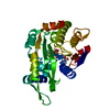
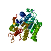
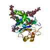
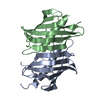
- PDB-3sih: The X-ray crystal structure of poly(ADP-ribose) glycohydrolase (P... -
+ Open data

- Basic information
| Entry | Database: PDB / ID: 3sih | ||||||
|---|---|---|---|---|---|---|---|
| Title | The X-ray crystal structure of poly(ADP-ribose) glycohydrolase (PARG) from Thermomonospora curvata | ||||||
 Components Components | poly(ADP-ribose) glycohydrolase | ||||||
 Keywords Keywords | HYDROLASE / poly ADP-ribose | ||||||
| Function / homology | Conserved hypothetical protein CHP02452 / Microbial-type PARG, catalytic domain / Microbial-type PARG, catalytic domain / Leucine Aminopeptidase, subunit E, domain 1 / Leucine Aminopeptidase, subunit E; domain 1 / Macro domain-like / 3-Layer(aba) Sandwich / Alpha Beta / Microbial-type PARG catalytic domain-containing protein Function and homology information Function and homology information | ||||||
| Biological species |  Thermomonospora curvata (bacteria) Thermomonospora curvata (bacteria) | ||||||
| Method |  X-RAY DIFFRACTION / SYNCHROTRON / SAD / Resolution: 1.5 Å X-RAY DIFFRACTION / SYNCHROTRON / SAD / Resolution: 1.5 Å | ||||||
 Authors Authors | Dunstan, M.S. / Leys, D. | ||||||
 Citation Citation | Journal: Nature / Year: 2011 Title: The structure and catalytic mechanism of a poly(ADP-ribose) glycohydrolase. Authors: Slade, D. / Dunstan, M.S. / Barkauskaite, E. / Weston, R. / Lafite, P. / Dixon, N. / Ahel, M. / Leys, D. / Ahel, I. | ||||||
| History |
|
- Structure visualization
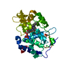
| Structure viewer | Molecule: MolmilJmol/JSmol |
|---|
- Downloads & links
-Download
| PDBx/mmCIF format | 3sih.cif.gz | 120.2 KB | Display | PDBx/mmCIF format |
|---|---|---|---|---|
| PDB format | pdb3sih.ent.gz | 93.8 KB | Display | PDB format |
| PDBx/mmJSON format | 3sih.json.gz | Tree view | PDBx/mmJSON format | |
| Others |  Other downloads Other downloads |
-Validation report
| Arichive directory | https://data.pdbj.org/pub/pdb/validation_reports/si/3sihftp://data.pdbj.org/pub/pdb/validation_reports/si/3sih | HTTPS FTP |
|---|
-Related structure data


-Links
 PDBj
PDBj- Assembly
| Deposited unit | 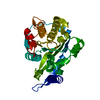
| ||||||||
|---|---|---|---|---|---|---|---|---|---|
| 1 |
| ||||||||
| Unit cell |
|
-Components
| #1: Protein | Mass: 29763.688 Da / Num. of mol.: 1 Source method: isolated from a genetically manipulated source Source: (gene. exp.) Thermomonospora curvata (bacteria) / Strain: ATCC 19995 / DSM 43183 / JCM 3096 / NCIMB 10081 / Gene: Tcur_1721 / Production host: References: UniProt: D1AC29, poly(ADP-ribose) glycohydrolase |
|---|---|
| #2: Water | ChemComp-HOH /  Mass: 18.015 Da / Num. of mol.: 226 / Source method: isolated from a natural source / Formula: H2O Mass: 18.015 Da / Num. of mol.: 226 / Source method: isolated from a natural source / Formula: H2O |
-Experimental details
-Experiment
| Experiment | Method: X-RAY DIFFRACTION / Number of used crystals: 1 |
|---|
- Sample preparation
| Crystal | Density Matthews: 1.96 Å3/Da / Density % sol: 37.36 % |
|---|---|
| Crystal grow | Method: sitting drop, vapor diffusion / pH: 9 Details: 10% PEG6000, 0.1 M Bicine, pH 9.0, SITTING DROP, VAPOR DIFFUSION |
-Data collection
| Diffraction | Mean temperature: 100 K |
|---|---|
| Diffraction source | Source: SYNCHROTRON / Site: Diamond  / Beamline: I04 / Wavelength: 0.976 / Beamline: I04 / Wavelength: 0.976 |
| Detector | Type: ADSC QUANTUM 315 / Detector: CCD / Date: Feb 24, 2011 |
| Radiation | Monochromator: Graphite / Protocol: SINGLE WAVELENGTH / Monochromatic (M) / Laue (L): M / Scattering type: x-ray |
| Radiation wavelength | Wavelength: 0.976 Å / Relative weight: 1 |
| Reflection | Resolution: 1.5→40.803 Å / Num. all: 38375 / Num. obs: 37023 / % possible obs: 96.5 % |
- Processing
| Software |
| ||||||||||||||||||||||||||||||||||||||||||||||||||||||||||||||||||||||||||||||||||||||||||||||||||
|---|---|---|---|---|---|---|---|---|---|---|---|---|---|---|---|---|---|---|---|---|---|---|---|---|---|---|---|---|---|---|---|---|---|---|---|---|---|---|---|---|---|---|---|---|---|---|---|---|---|---|---|---|---|---|---|---|---|---|---|---|---|---|---|---|---|---|---|---|---|---|---|---|---|---|---|---|---|---|---|---|---|---|---|---|---|---|---|---|---|---|---|---|---|---|---|---|---|---|---|
| Refinement | Method to determine structure: SAD / Resolution: 1.5→33.156 Å / Occupancy max: 1 / Occupancy min: 1 / FOM work R set: 0.853 / SU ML: 0.16 / σ(F): 2 / Phase error: 21.49 / Stereochemistry target values: ML
| ||||||||||||||||||||||||||||||||||||||||||||||||||||||||||||||||||||||||||||||||||||||||||||||||||
| Solvent computation | Shrinkage radii: 0.9 Å / VDW probe radii: 1.11 Å / Solvent model: FLAT BULK SOLVENT MODEL / Bsol: 51.031 Å2 / ksol: 0.357 e/Å3 | ||||||||||||||||||||||||||||||||||||||||||||||||||||||||||||||||||||||||||||||||||||||||||||||||||
| Displacement parameters | Biso max: 113.54 Å2 / Biso mean: 28.2533 Å2 / Biso min: 7.62 Å2
| ||||||||||||||||||||||||||||||||||||||||||||||||||||||||||||||||||||||||||||||||||||||||||||||||||
| Refine analyze | Luzzati sigma a obs: 0.16 Å | ||||||||||||||||||||||||||||||||||||||||||||||||||||||||||||||||||||||||||||||||||||||||||||||||||
| Refinement step | Cycle: LAST / Resolution: 1.5→33.156 Å
| ||||||||||||||||||||||||||||||||||||||||||||||||||||||||||||||||||||||||||||||||||||||||||||||||||
| Refine LS restraints |
| ||||||||||||||||||||||||||||||||||||||||||||||||||||||||||||||||||||||||||||||||||||||||||||||||||
| LS refinement shell | Refine-ID: X-RAY DIFFRACTION / Total num. of bins used: 13
| ||||||||||||||||||||||||||||||||||||||||||||||||||||||||||||||||||||||||||||||||||||||||||||||||||
| Refinement TLS params. | Method: refined / Origin x: 35.1075 Å / Origin y: 29.5077 Å / Origin z: 13.8455 Å
| ||||||||||||||||||||||||||||||||||||||||||||||||||||||||||||||||||||||||||||||||||||||||||||||||||
| Refinement TLS group |
|
