 Movie
Movie Controller

[English] 日本語
 Yorodumi
- PDB-3sce: Structure of the Thioalkalivibrio nitratireducens cytochrome c ni... -
+ Open data

- Basic information
| Entry | Database: PDB / ID: 3sce | ||||||
|---|---|---|---|---|---|---|---|
| Title | Structure of the Thioalkalivibrio nitratireducens cytochrome c nitrite reductase with a covalent bond between the CE1 atom of Tyr303 and the CG atom of Gln360 (TvNiRb) | ||||||
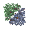
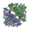
 Components Components | Eight-heme nitrite reductase | ||||||
 Keywords Keywords | OXIDOREDUCTASE / eight hemes c / nitrite reductase / Tyr-Gln and Tyr-Cys bonds | ||||||
| Function / homology |  Function and homology information Function and homology informationanaerobic respiration, using ammonium as electron donor / nitrite reductase (cytochrome; ammonia-forming) / nitrite reductase (cytochrome, ammonia-forming) activity / anaerobic electron transport chain / nitrate assimilation / outer membrane-bounded periplasmic space / heme binding / calcium ion binding Similarity search - Function | ||||||
| Biological species |  Thioalkalivibrio nitratireducens (bacteria) Thioalkalivibrio nitratireducens (bacteria) | ||||||
| Method |  X-RAY DIFFRACTION / SYNCHROTRON / MOLECULAR REPLACEMENT / Resolution: 1.45 Å X-RAY DIFFRACTION / SYNCHROTRON / MOLECULAR REPLACEMENT / Resolution: 1.45 Å | ||||||
 Authors Authors | Trofimov, A.A. / Polyakov, K.M. / Boyko, K.M. / Tikhonova, T.V. / Popov, V.O. | ||||||
 Citation Citation | Journal: Acta Crystallogr.,Sect.D / Year: 2012 Title: Covalent modifications of the catalytic tyrosine in octahaem cytochrome c nitrite reductase and their effect on the enzyme activity. Authors: Trofimov, A.A. / Polyakov, K.M. / Tikhonova, T.V. / Tikhonov, A.V. / Safonova, T.N. / Boyko, K.M. / Dorovatovskii, P.V. / Popov, V.O. | ||||||
| History |
|
- Structure visualization
| Structure viewer | Molecule: MolmilJmol/JSmol |
|---|
- Downloads & links
-Download
| PDBx/mmCIF format | 3sce.cif.gz | 519.4 KB | Display | PDBx/mmCIF format |
|---|---|---|---|---|
| PDB format | pdb3sce.ent.gz | 426.3 KB | Display | PDB format |
| PDBx/mmJSON format | 3sce.json.gz | Tree view | PDBx/mmJSON format | |
| Others |  Other downloads Other downloads |
-Validation report
| Arichive directory | https://data.pdbj.org/pub/pdb/validation_reports/sc/3sceftp://data.pdbj.org/pub/pdb/validation_reports/sc/3sce | HTTPS FTP |
|---|
-Related structure data
| Related structure data |  3lg1C  3lgqC  3rkhC 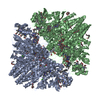 3uu9C 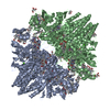 2ot4S S: Starting model for refinement C: citing same article ( |
|---|---|
| Similar structure data |

-Links
 PDBj
PDBj


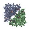
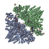
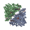
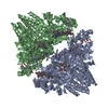
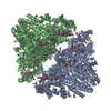
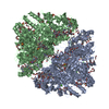
- Assembly
| Deposited unit | 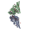
| |||||||||||||||||||||||||||
|---|---|---|---|---|---|---|---|---|---|---|---|---|---|---|---|---|---|---|---|---|---|---|---|---|---|---|---|---|
| 1 | 
| |||||||||||||||||||||||||||
| Unit cell |
| |||||||||||||||||||||||||||
| Components on special symmetry positions |
|
-Components
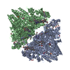
-Protein , 1 types, 2 molecules AB
| #1: Protein | Mass: 58834.641 Da / Num. of mol.: 2 / Fragment: UNP residues 33-551 / Source method: isolated from a natural source Source: (natural) Thioalkalivibrio nitratireducens (bacteria)Strain: ALEN 2 References: UniProt: Q5F2I3, UniProt: L0DSL2*PLUS, Oxidoreductases; Acting on other nitrogenous compounds as donors; With a cytochrome as acceptor |
|---|
-Non-polymers , 7 types, 1233 molecules 


| #2: Chemical | ChemComp-HEC /  Mass: 618.503 Da / Num. of mol.: 16 / Source method: obtained synthetically / Formula: C34H34FeN4O4 Mass: 618.503 Da / Num. of mol.: 16 / Source method: obtained synthetically / Formula: C34H34FeN4O4#3: Chemical | ChemComp-PO4 /  Mass: 94.971 Da / Num. of mol.: 4 / Source method: obtained synthetically / Formula: PO4 Mass: 94.971 Da / Num. of mol.: 4 / Source method: obtained synthetically / Formula: PO4#4: Chemical | ChemComp-CA /  Mass: 40.078 Da / Num. of mol.: 4 / Source method: obtained synthetically / Formula: Ca Mass: 40.078 Da / Num. of mol.: 4 / Source method: obtained synthetically / Formula: Ca#5: Chemical | ChemComp-PG4 /  Mass: 194.226 Da / Num. of mol.: 16 / Source method: obtained synthetically / Formula: C8H18O5 / Comment: precipitant*YM Mass: 194.226 Da / Num. of mol.: 16 / Source method: obtained synthetically / Formula: C8H18O5 / Comment: precipitant*YM#6: Chemical | ChemComp-PG6 /  Mass: 266.331 Da / Num. of mol.: 5 / Source method: obtained synthetically / Formula: C12H26O6 Mass: 266.331 Da / Num. of mol.: 5 / Source method: obtained synthetically / Formula: C12H26O6#7: Chemical |  Mass: 22.990 Da / Num. of mol.: 2 / Source method: obtained synthetically / Formula: Na Mass: 22.990 Da / Num. of mol.: 2 / Source method: obtained synthetically / Formula: Na#8: Water | ChemComp-HOH / | Mass: 18.015 Da / Num. of mol.: 1186 / Source method: isolated from a natural source / Formula: H2O |
|---|
-Details
| Has protein modification | Y |
|---|
-Experimental details
-Experiment
| Experiment | Method: X-RAY DIFFRACTION / Number of used crystals: 1 |
|---|
- Sample preparation
| Crystal | Density Matthews: 5.09 Å3/Da / Density % sol: 75.84 % |
|---|---|
| Crystal grow | Temperature: 278 K / Method: vapor diffusion, hanging drop / pH: 8.7 Details: Protein solution (2.5 uL): 9.3 mg/mL TvNiRb, 0.1 M potassium phosphate, pH 7.0, Reservoir solution (2.5 uL): 0.2 M tri-sodium citrate dihydrate, 0.1 M Tris hydrochloride, pH8.5, 30% v/v ...Details: Protein solution (2.5 uL): 9.3 mg/mL TvNiRb, 0.1 M potassium phosphate, pH 7.0, Reservoir solution (2.5 uL): 0.2 M tri-sodium citrate dihydrate, 0.1 M Tris hydrochloride, pH8.5, 30% v/v PEG400 , VAPOR DIFFUSION, HANGING DROP, temperature 278.0 K |
-Data collection
| Diffraction | Mean temperature: 100 K |
|---|---|
| Diffraction source | Source: SYNCHROTRON / Site: SPring-8  / Beamline: BL41XU / Wavelength: 0.75 / Beamline: BL41XU / Wavelength: 0.75 |
| Detector | Type: MAR CCD 165 mm / Detector: CCD |
| Radiation | Protocol: SINGLE WAVELENGTH / Monochromatic (M) / Laue (L): M / Scattering type: x-ray |
| Radiation wavelength | Wavelength: 0.75 Å / Relative weight: 1 |
| Reflection | Resolution: 1.45→136.472 Å / Num. obs: 464187 / % possible obs: 99.9 % / Biso Wilson estimate: 21.2 Å2 / Rmerge(I) obs: 0.11 / Net I/σ(I): 11.1 |
| Reflection shell | Resolution: 1.45→1.6 Å / Rmerge(I) obs: 0.544 / Mean I/σ(I) obs: 3.4 / % possible all: 99.9 |
- Processing
| Software |
| ||||||||||||||||||||||||||||||||||||||||||||||||||||||||||||||||||||||||||||||||||||||||||||||||||||
|---|---|---|---|---|---|---|---|---|---|---|---|---|---|---|---|---|---|---|---|---|---|---|---|---|---|---|---|---|---|---|---|---|---|---|---|---|---|---|---|---|---|---|---|---|---|---|---|---|---|---|---|---|---|---|---|---|---|---|---|---|---|---|---|---|---|---|---|---|---|---|---|---|---|---|---|---|---|---|---|---|---|---|---|---|---|---|---|---|---|---|---|---|---|---|---|---|---|---|---|---|---|
| Refinement | Method to determine structure: MOLECULAR REPLACEMENT Starting model: PDB ENTRY 2OT4 Resolution: 1.45→100 Å / Cor.coef. Fo:Fc: 0.979 / Cor.coef. Fo:Fc free: 0.975 / SU B: 1.146 / SU ML: 0.02 / Cross valid method: THROUGHOUT / ESU R: 0.034 / ESU R Free: 0.034 / Stereochemistry target values: MAXIMUM LIKELIHOOD Details: HYDROGENS HAVE BEEN ADDED IN THE RIDING POSITIONS U VALUES : REFINED INDIVIDUALLY
| ||||||||||||||||||||||||||||||||||||||||||||||||||||||||||||||||||||||||||||||||||||||||||||||||||||
| Solvent computation | Ion probe radii: 0.8 Å / Shrinkage radii: 0.8 Å / VDW probe radii: 1.4 Å / Solvent model: BABINET MODEL WITH MASK | ||||||||||||||||||||||||||||||||||||||||||||||||||||||||||||||||||||||||||||||||||||||||||||||||||||
| Displacement parameters | Biso mean: 14.342 Å2 | ||||||||||||||||||||||||||||||||||||||||||||||||||||||||||||||||||||||||||||||||||||||||||||||||||||
| Refine analyze | Luzzati coordinate error obs: 0.139 Å | ||||||||||||||||||||||||||||||||||||||||||||||||||||||||||||||||||||||||||||||||||||||||||||||||||||
| Refinement step | Cycle: LAST / Resolution: 1.45→100 Å
| ||||||||||||||||||||||||||||||||||||||||||||||||||||||||||||||||||||||||||||||||||||||||||||||||||||
| Refine LS restraints |
| ||||||||||||||||||||||||||||||||||||||||||||||||||||||||||||||||||||||||||||||||||||||||||||||||||||
| LS refinement shell | Resolution: 1.45→1.488 Å / Total num. of bins used: 20
|
