 Movie
Movie Controller

[English] 日本語
 Yorodumi
- PDB-2ot4: Structure of a hexameric multiheme c nitrite reductase from the e... -
+ Open data

- Basic information
| Entry | Database: PDB / ID: 2ot4 | ||||||
|---|---|---|---|---|---|---|---|
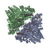
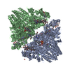
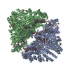
| Title | Structure of a hexameric multiheme c nitrite reductase from the extremophile bacterium Thiolkalivibrio nitratireducens | ||||||
 Components Components | Eight-heme nitrite reductase | ||||||
 Keywords Keywords | OXIDOREDUCTASE / cytochrome c nitrite reductase / NrfA / sulfite reductase | ||||||
| Function / homology |  Function and homology information Function and homology informationanaerobic respiration, using ammonium as electron donor / nitrite reductase (cytochrome; ammonia-forming) / nitrite reductase (cytochrome, ammonia-forming) activity / anaerobic electron transport chain / nitrate assimilation / outer membrane-bounded periplasmic space / heme binding / calcium ion binding Similarity search - Function | ||||||
| Biological species |  Thioalkalivibrio nitratireducens (bacteria) Thioalkalivibrio nitratireducens (bacteria) | ||||||
| Method |  X-RAY DIFFRACTION / SYNCHROTRON / SAD / Resolution: 1.5 Å X-RAY DIFFRACTION / SYNCHROTRON / SAD / Resolution: 1.5 Å | ||||||
 Authors Authors | Polyakov, K.M. / Boyko, K.M. / Slutsky, A. / Tikhonova, T.V. / Antipov, A.N. / Zvyagilskaya, R.A. / Popov, A.N. / Lamzin, V.S. / Bourenkov, G.P. / Popov, V.O. | ||||||
 Citation Citation | Journal: J.Mol.Biol. / Year: 2009 Title: High-resolution structural analysis of a novel octaheme cytochrome c nitrite reductase from the haloalkaliphilic bacterium Thioalkalivibrio nitratireducens. Authors: Polyakov, K.M. / Boyko, K.M. / Tikhonova, T.V. / Slutsky, A. / Antipov, A.N. / Zvyagilskaya, R.A. / Popov, A.N. / Bourenkov, G.P. / Lamzin, V.S. / Popov, V.O. #1: Journal: Acta Crystallogr.,Sect.F / Year: 2006Title: Crystallization and preliminary X-ray analysis of cytochrome c nitrite reductase from Thialkalivibrio nitraterecense. Authors: Boyko, K.M. / Polyakov, K.M. / Tikhonova, T.V. / Slutsky, A. / Antipov, A.N. / Zvyagilskaya, R.A. / Bourenkov, G.P. / Popov, A.N. / Lamzin, V.S. / Popov, V.O. #2: Journal: BIOCHIM.BIOPHYS.ACTA / Year: 2006Title: Molecular and catalytic properties of a novel cytochrome c nitrite reductase from nitrate-reducing haloalkaliphic sulfur-oxidixing bacterium Thioalkalivibrio nitratireducens. Authors: Tikhonova, T.V. / Slutsky, A. / Antipov, A.N. / Boyko, K.M. / Polyakov, K.M. / Sorokin, D.Y. / Zvyagilskaya, R.A. / Popov, V.O. #3: Journal: Acta Crystallogr.,Sect.D / Year: 2010Title: Structures of complexes of octahaem cytochrome c nitrite reductase from Thioalkalivibrio nitratireducens with sulfite and cyanide. Authors: Trofimov, A.A. / Polyakov, K.M. / Boyko, K.M. / Tikhonova, T.V. / Safonova, T.N. / Tikhonov, A.V. / Popov, A.N. / Popov, V.O. #4: Journal: Crystallography Reports / Year: 2010Title: Structure of octaheme cytochrome c nitrite reductase from Thioalkalivibrio nitratireducens in a complex with phosphate Authors: Trofimov, A.A. / Polyakov, K.M. / Boiko, K.M. / Filimonenkov, A.A. / Dorovatovskii, P.V. / Tikhonova, T.V. / Popov, V.O. / Kovalchuk, M.V. | ||||||
| History |
| ||||||
| Remark 400 | COMPOUND UNUSUAL COVALENT BOND WAS DETECTED IN THE ACTIVE SITE OF THE ENZYME BETWEEN S ATOM OF ...COMPOUND UNUSUAL COVALENT BOND WAS DETECTED IN THE ACTIVE SITE OF THE ENZYME BETWEEN S ATOM OF CYS305 AND ORTHO-POSITION OF TYR303. |
- Structure visualization
| Structure viewer | Molecule: MolmilJmol/JSmol |
|---|
- Downloads & links
-Download
| PDBx/mmCIF format | 2ot4.cif.gz | 856.6 KB | Display | PDBx/mmCIF format |
|---|---|---|---|---|
| PDB format | pdb2ot4.ent.gz | 718 KB | Display | PDB format |
| PDBx/mmJSON format | 2ot4.json.gz | Tree view | PDBx/mmJSON format | |
| Others |  Other downloads Other downloads |
-Validation report
| Arichive directory | https://data.pdbj.org/pub/pdb/validation_reports/ot/2ot4ftp://data.pdbj.org/pub/pdb/validation_reports/ot/2ot4 | HTTPS FTP |
|---|
-Related structure data


-Links
 PDBj
PDBj


- Assembly
| Deposited unit | 
| |||||||||||||||
|---|---|---|---|---|---|---|---|---|---|---|---|---|---|---|---|---|
| 1 | 
| |||||||||||||||
| Unit cell |
| |||||||||||||||
| Components on special symmetry positions |
| |||||||||||||||
| Details | The biological assembly is a hexamer generated from the dimer in the asymmetric unit(chain A and chain B) by the following operations: z-1/2, -x+1/2, -y+1 and -y+1/2, -z+1, x+1/2. |
-Components
-Protein , 1 types, 2 molecules AB
| #1: Protein | Mass: 59492.293 Da / Num. of mol.: 2 / Source method: isolated from a natural source Source: (natural) Thioalkalivibrio nitratireducens (bacteria)Strain: ALEN 2 / References: UniProt: Q5F2I3, UniProt: L0DSL2*PLUS |
|---|
-Non-polymers , 6 types, 1243 molecules 


| #2: Chemical |  Mass: 40.078 Da / Num. of mol.: 2 / Source method: obtained synthetically / Formula: Ca Mass: 40.078 Da / Num. of mol.: 2 / Source method: obtained synthetically / Formula: Ca#3: Chemical | ChemComp-HEC /  Mass: 618.503 Da / Num. of mol.: 16 / Source method: obtained synthetically / Formula: C34H34FeN4O4 Mass: 618.503 Da / Num. of mol.: 16 / Source method: obtained synthetically / Formula: C34H34FeN4O4#4: Chemical | ChemComp-MRD / (  Mass: 118.174 Da / Num. of mol.: 4 / Source method: obtained synthetically / Formula: C6H14O2 / Comment: precipitant*YM Mass: 118.174 Da / Num. of mol.: 4 / Source method: obtained synthetically / Formula: C6H14O2 / Comment: precipitant*YM#5: Chemical |  Mass: 118.174 Da / Num. of mol.: 3 / Source method: obtained synthetically / Formula: C6H14O2 / Comment: precipitant*YM Mass: 118.174 Da / Num. of mol.: 3 / Source method: obtained synthetically / Formula: C6H14O2 / Comment: precipitant*YM#6: Chemical | ChemComp-CIT / |  Mass: 192.124 Da / Num. of mol.: 1 / Source method: obtained synthetically / Formula: C6H8O7 Mass: 192.124 Da / Num. of mol.: 1 / Source method: obtained synthetically / Formula: C6H8O7#7: Water | ChemComp-HOH / | Mass: 18.015 Da / Num. of mol.: 1217 / Source method: isolated from a natural source / Formula: H2O |
|---|
-Details
| Has protein modification | Y |
|---|
-Experimental details
-Experiment
| Experiment | Method: X-RAY DIFFRACTION / Number of used crystals: 1 |
|---|
- Sample preparation
| Crystal grow | Temperature: 278 K / Method: vapor diffusion, hanging drop / pH: 5.6 Details: The drop contained 5mkl of enzyme solution (14.5 mg/ml) in 0.005 M Tris-borat buffer (pH 8.7) and 5 mkl reservoir solution. The reservoir solution contained 26.3% MPD, 0.175 M ammonium ...Details: The drop contained 5mkl of enzyme solution (14.5 mg/ml) in 0.005 M Tris-borat buffer (pH 8.7) and 5 mkl reservoir solution. The reservoir solution contained 26.3% MPD, 0.175 M ammonium acetate in 0.09 M sodium citrate buffer (pH 5.6), VAPOR DIFFUSION, HANGING DROP, temperature 278K |
|---|
-Data collection
| Diffraction | Mean temperature: 100 K | |||||||||
|---|---|---|---|---|---|---|---|---|---|---|
| Diffraction source | Source: SYNCHROTRON / Site: EMBL/DESY, HAMBURG  / Beamline: BW7A / Wavelength: 0.96, 1.7375 / Beamline: BW7A / Wavelength: 0.96, 1.7375 | |||||||||
| Detector | Type: MAR CCD 165 mm / Detector: CCD | |||||||||
| Radiation | Protocol: SINGLE WAVELENGTH / Monochromatic (M) / Laue (L): M / Scattering type: x-ray | |||||||||
| Radiation wavelength |
| |||||||||
| Reflection | Resolution: 1.5→30 Å / Num. obs: 373791 / % possible obs: 97.4 % / Biso Wilson estimate: 17.1 Å2 / Rmerge(I) obs: 0.036 / Χ2: 1.241 / Net I/σ(I): 17.1 | |||||||||
| Reflection shell | Resolution: 1.5→1.52 Å / Rmerge(I) obs: 0.476 / Num. unique all: 12460 / Χ2: 1.764 / % possible all: 98.3 |
- Processing
| Software |
| |||||||||||||||||||||||||||||||||||||||||||||||||||||||||||||||||||||||||||||||||||||||||||||||||||||||||||||||||||||||||||||||||||||||||||||||||
|---|---|---|---|---|---|---|---|---|---|---|---|---|---|---|---|---|---|---|---|---|---|---|---|---|---|---|---|---|---|---|---|---|---|---|---|---|---|---|---|---|---|---|---|---|---|---|---|---|---|---|---|---|---|---|---|---|---|---|---|---|---|---|---|---|---|---|---|---|---|---|---|---|---|---|---|---|---|---|---|---|---|---|---|---|---|---|---|---|---|---|---|---|---|---|---|---|---|---|---|---|---|---|---|---|---|---|---|---|---|---|---|---|---|---|---|---|---|---|---|---|---|---|---|---|---|---|---|---|---|---|---|---|---|---|---|---|---|---|---|---|---|---|---|---|---|---|
| Refinement | Method to determine structure: SAD / Resolution: 1.5→24.04 Å / Cor.coef. Fo:Fc: 0.98 / Cor.coef. Fo:Fc free: 0.976 / SU B: 1.22 / SU ML: 0.021 / Cross valid method: THROUGHOUT / σ(F): 0 / ESU R: 0.038 / ESU R Free: 0.036 / Stereochemistry target values: MAXIMUM LIKELIHOOD / Details: HYDROGENS HAVE BEEN ADDED IN THE RIDING POSITIONS
| |||||||||||||||||||||||||||||||||||||||||||||||||||||||||||||||||||||||||||||||||||||||||||||||||||||||||||||||||||||||||||||||||||||||||||||||||
| Solvent computation | Ion probe radii: 0.8 Å / Shrinkage radii: 0.8 Å / VDW probe radii: 1.2 Å / Solvent model: BABINET MODEL WITH MASK | |||||||||||||||||||||||||||||||||||||||||||||||||||||||||||||||||||||||||||||||||||||||||||||||||||||||||||||||||||||||||||||||||||||||||||||||||
| Displacement parameters | Biso mean: 15.294 Å2 | |||||||||||||||||||||||||||||||||||||||||||||||||||||||||||||||||||||||||||||||||||||||||||||||||||||||||||||||||||||||||||||||||||||||||||||||||
| Refine analyze | Luzzati coordinate error obs: 0.137 Å | |||||||||||||||||||||||||||||||||||||||||||||||||||||||||||||||||||||||||||||||||||||||||||||||||||||||||||||||||||||||||||||||||||||||||||||||||
| Refinement step | Cycle: LAST / Resolution: 1.5→24.04 Å
| |||||||||||||||||||||||||||||||||||||||||||||||||||||||||||||||||||||||||||||||||||||||||||||||||||||||||||||||||||||||||||||||||||||||||||||||||
| Refine LS restraints |
| |||||||||||||||||||||||||||||||||||||||||||||||||||||||||||||||||||||||||||||||||||||||||||||||||||||||||||||||||||||||||||||||||||||||||||||||||
| LS refinement shell | Resolution: 1.5→1.539 Å / Total num. of bins used: 20
|
