- PDB-3s7x: Unassembled Washington University Polyomavirus VP1 Pentamer R198K... -
+
Open data
ID or keywords:
Loading...
-
Basic information
Entry
Database: PDB / ID: 3s7x
Title
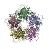
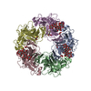
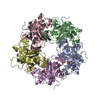
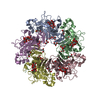
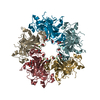
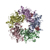
Unassembled Washington University Polyomavirus VP1 Pentamer R198K Mutant
Components
Major capsid protein VP1
Keywords
VIRAL PROTEIN / jelly-roll fold / antiparallel beta sandwich / major capsid protein
Function / homology
Function and homology information
T=7 icosahedral viral capsid / endocytosis involved in viral entry into host cell / virion attachment to host cell / host cell nucleus / structural molecule activity Similarity search - Function
Capsid protein VP1,Polyomavirus / Polyomavirus Vp1; Chain A / Capsid protein VP1,Polyomavirus / Polyomavirus capsid protein VP1 superfamily / Polyomavirus coat protein / Double-stranded DNA virus, group I, capsid / Sandwich / Mainly Beta Similarity search - Domain/homology
#221 - May 2018 Human Papillomavirus and Vaccines similarity (1)
#200 - Aug 2016 Quasisymmetry in Icosahedral Viruses similarity (5)
-
Assembly
Deposited unit
A: Major capsid protein VP1 B: Major capsid protein VP1 C: Major capsid protein VP1 D: Major capsid protein VP1 E: Major capsid protein VP1 hetero molecules
Mass: 22.990 Da / Num. of mol.: 5 / Source method: obtained synthetically / Formula: Na
Has protein modification
Y
Sequence details
THE R198K MUTATION WAS INTRODUCED INTO THE WILD-TYPE PROTEIN TO REMOVE A NON-CANONICAL THROMBIN ...THE R198K MUTATION WAS INTRODUCED INTO THE WILD-TYPE PROTEIN TO REMOVE A NON-CANONICAL THROMBIN CLEAVAGE SITE TO OBTAIN A HOMOGENEOUS SAMPLE FOR CRYSTALLIZATION
-
Experimental details
-
Experiment
Experiment
Method: X-RAY DIFFRACTION / Number of used crystals: 1
-
Sample preparation
Crystal
Density Matthews: 3.04 Å3/Da / Density % sol: 59.48 %
Crystal grow
Temperature: 282 K / Method: vapor diffusion, sitting drop / pH: 7.5 Details: 0.2 M trisodium citrate, 0.1 M HEPES pH 7.5, 20 % (v/v) isopropanol, VAPOR DIFFUSION, SITTING DROP, temperature 282K
Protocol: SINGLE WAVELENGTH / Monochromatic (M) / Laue (L): M / Scattering type: x-ray
Radiation wavelength
Wavelength: 0.933 Å / Relative weight: 1
Reflection
Resolution: 2.9→40 Å / Num. all: 40144 / Num. obs: 39647 / % possible obs: 98.8 % / Observed criterion σ(I): -3 / Rmerge(I) obs: 0.096 / Net I/σ(I): 11.1
Reflection shell
Resolution: 2.9→2.98 Å / Rmerge(I) obs: 0.578 / Mean I/σ(I) obs: 1.8 / % possible all: 99.4
-
Processing
Software
Name
Version
Classification
MxCuBE
datacollection
PHASER
phasing
REFMAC
5.5.0102
refinement
XDS
datareduction
XSCALE
datascaling
Refinement
Method to determine structure: MOLECULAR REPLACEMENT / Resolution: 2.9→43.36 Å / Cor.coef. Fo:Fc: 0.921 / Cor.coef. Fo:Fc free: 0.906 / SU B: 17.497 / SU ML: 0.315 / Cross valid method: THROUGHOUT / ESU R Free: 0.394 / Stereochemistry target values: MAXIMUM LIKELIHOOD / Details: HYDROGENS HAVE BEEN ADDED IN THE RIDING POSITIONS
Rfactor
Num. reflection
% reflection
Selection details
Rfree
0.25296
3038
7.6 %
RANDOM
Rwork
0.234
-
-
-
obs
0.23545
36689
98.73 %
-
all
-
39647
-
-
Solvent computation
Ion probe radii: 0.8 Å / Shrinkage radii: 0.8 Å / VDW probe radii: 1.2 Å / Solvent model: MASK
Displacement parameters
Biso mean: 60.169 Å2
Baniso -1
Baniso -2
Baniso -3
1-
1.68 Å2
0 Å2
0 Å2
2-
-
1.68 Å2
0 Å2
3-
-
-
-3.36 Å2
Refinement step
Cycle: LAST / Resolution: 2.9→43.36 Å
Protein
Nucleic acid
Ligand
Solvent
Total
Num. atoms
9935
0
40
0
9975
Refine LS restraints
Refine-ID
Type
Dev ideal
Dev ideal target
Number
X-RAY DIFFRACTION
r_bond_refined_d
0.008
0.022
10209
X-RAY DIFFRACTION
r_bond_other_d
X-RAY DIFFRACTION
r_angle_refined_deg
1.197
1.969
13942
X-RAY DIFFRACTION
r_angle_other_deg
X-RAY DIFFRACTION
r_dihedral_angle_1_deg
6.427
5
1291
X-RAY DIFFRACTION
r_dihedral_angle_2_deg
35.883
23.918
416
X-RAY DIFFRACTION
r_dihedral_angle_3_deg
17.367
15
1556
X-RAY DIFFRACTION
r_dihedral_angle_4_deg
21.932
15
60
X-RAY DIFFRACTION
r_chiral_restr
0.082
0.2
1593
X-RAY DIFFRACTION
r_gen_planes_refined
0.004
0.022
7794
X-RAY DIFFRACTION
r_gen_planes_other
X-RAY DIFFRACTION
r_nbd_refined
X-RAY DIFFRACTION
r_nbd_other
X-RAY DIFFRACTION
r_nbtor_refined
X-RAY DIFFRACTION
r_nbtor_other
X-RAY DIFFRACTION
r_xyhbond_nbd_refined
X-RAY DIFFRACTION
r_xyhbond_nbd_other
X-RAY DIFFRACTION
r_metal_ion_refined
X-RAY DIFFRACTION
r_metal_ion_other
X-RAY DIFFRACTION
r_symmetry_vdw_refined
X-RAY DIFFRACTION
r_symmetry_vdw_other
X-RAY DIFFRACTION
r_symmetry_hbond_refined
X-RAY DIFFRACTION
r_symmetry_hbond_other
X-RAY DIFFRACTION
r_symmetry_metal_ion_refined
X-RAY DIFFRACTION
r_symmetry_metal_ion_other
X-RAY DIFFRACTION
r_mcbond_it
1.637
2
6488
X-RAY DIFFRACTION
r_mcbond_other
X-RAY DIFFRACTION
r_mcangle_it
3.008
4
10535
X-RAY DIFFRACTION
r_scbond_it
1.896
2
3719
X-RAY DIFFRACTION
r_scangle_it
3.354
4
3407
X-RAY DIFFRACTION
r_rigid_bond_restr
X-RAY DIFFRACTION
r_sphericity_free
X-RAY DIFFRACTION
r_sphericity_bonded
Refine LS restraints NCS
Ens-ID: 1 / Refine-ID: X-RAY DIFFRACTION
Dom-ID
Auth asym-ID
Number
Type
Rms dev position (Å)
Weight position
1
A
960
tightpositional
0.02
0.05
2
B
960
tightpositional
0.02
0.05
3
C
960
tightpositional
0.02
0.05
4
D
960
tightpositional
0.02
0.05
5
E
960
tightpositional
0.02
0.05
1
A
885
loosepositional
0.02
5
2
B
885
loosepositional
0.02
5
3
C
885
loosepositional
0.02
5
4
D
885
loosepositional
0.02
5
5
E
885
loosepositional
0.02
5
1
A
960
tightthermal
0.04
0.5
2
B
960
tightthermal
0.04
0.5
3
C
960
tightthermal
0.04
0.5
4
D
960
tightthermal
0.04
0.5
5
E
960
tightthermal
0.04
0.5
1
A
885
loosethermal
0.03
10
2
B
885
loosethermal
0.03
10
3
C
885
loosethermal
0.03
10
4
D
885
loosethermal
0.03
10
5
E
885
loosethermal
0.03
10
LS refinement shell
Resolution: 2.897→2.972 Å / Total num. of bins used: 20
Rfactor
Num. reflection
% reflection
Rfree
0.349
215
-
Rwork
0.37
2649
-
obs
-
-
98.35 %
+
About Yorodumi
-
News
-
Feb 9, 2022. New format data for meta-information of EMDB entries
New format data for meta-information of EMDB entries
Version 3 of the EMDB header file is now the official format.
The previous official version 1.9 will be removed from the archive.
In the structure databanks used in Yorodumi, some data are registered as the other names, "COVID-19 virus" and "2019-nCoV". Here are the details of the virus and the list of structure data.
Jan 31, 2019. EMDB accession codes are about to change! (news from PDBe EMDB page)
EMDB accession codes are about to change! (news from PDBe EMDB page)
The allocation of 4 digits for EMDB accession codes will soon come to an end. Whilst these codes will remain in use, new EMDB accession codes will include an additional digit and will expand incrementally as the available range of codes is exhausted. The current 4-digit format prefixed with “EMD-” (i.e. EMD-XXXX) will advance to a 5-digit format (i.e. EMD-XXXXX), and so on. It is currently estimated that the 4-digit codes will be depleted around Spring 2019, at which point the 5-digit format will come into force.
The EM Navigator/Yorodumi systems omit the EMD- prefix.
Related info.:Q: What is EMD? / ID/Accession-code notation in Yorodumi/EM Navigator
Yorodumi is a browser for structure data from EMDB, PDB, SASBDB, etc.
This page is also the successor to EM Navigator detail page, and also detail information page/front-end page for Omokage search.
The word "yorodu" (or yorozu) is an old Japanese word meaning "ten thousand". "mi" (miru) is to see.
Related info.:EMDB / PDB / SASBDB / Comparison of 3 databanks / Yorodumi Search / Aug 31, 2016. New EM Navigator & Yorodumi / Yorodumi Papers / Jmol/JSmol / Function and homology information / Changes in new EM Navigator and Yorodumi
 Movie
Movie Controller

 Yorodumi
 Open data
 Basic information
 Components
 Keywords
 Function and homology information
 WU Polyomavirus
 X-RAY DIFFRACTION /
  Authors
 Citation
 Structure visualization
 Downloads & links
 Other downloads


 PDBj
PDBj


 Assembly
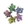


 Mass: 92.094 Da / Num. of mol.: 5 / Source method: obtained synthetically / Formula: C3H8O3
 Mass: 35.453 Da / Num. of mol.: 5 / Source method: obtained synthetically / Formula: Cl
 Mass: 22.990 Da / Num. of mol.: 5 / Source method: obtained synthetically / Formula: Na
 Sample preparation
 / Beamline: ID14-1 / Wavelength: 0.933 Å
 Processing