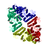
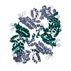
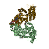
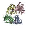
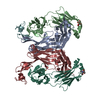
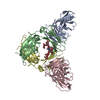
Entry Database : PDB / ID : 3s6hTitle Crystal structure of native mmNAGS/k N-acetylglutamate kinase / N-acetylglutamate synthase Keywords / Function / homology Function Domain/homology Component
/ / / / / / / / / / / / / / / / / / / / / / / / / / / / / / / Biological species Maricaulis maris (bacteria)Method / / / Resolution : 3.102 Å Authors Shi, D. / Li, Y. / Cabrera-Luque, J. / Jin, Z. / Yu, X. / Allewell, N.M. / Tuchman, M. Journal : Plos One / Year : 2011Title : A Novel N-acetylglutamate synthase architecture revealed by the crystal structure of the bifunctional enzyme from Maricaulis maris.Authors : Shi, D. / Li, Y. / Cabrera-Luque, J. / Jin, Z. / Yu, X. / Zhao, G. / Haskins, N. / Allewell, N.M. / Tuchman, M. History Deposition May 25, 2011 Deposition site / Processing site Revision 1.0 Apr 25, 2012 Provider / Type Revision 1.1 Sep 13, 2023 Group Data collection / Database references ... Data collection / Database references / Derived calculations / Refinement description Category chem_comp_atom / chem_comp_bond ... chem_comp_atom / chem_comp_bond / database_2 / pdbx_initial_refinement_model / struct_ref_seq_dif / struct_site Item _database_2.pdbx_DOI / _database_2.pdbx_database_accession ... _database_2.pdbx_DOI / _database_2.pdbx_database_accession / _struct_ref_seq_dif.details / _struct_site.pdbx_auth_asym_id / _struct_site.pdbx_auth_comp_id / _struct_site.pdbx_auth_seq_id
Show all Show less
 Movie
Movie Controller


 Open data
 Basic information
 Components
 Keywords
 Function and homology information
 Maricaulis maris (bacteria)
 X-RAY DIFFRACTION /
  Authors
 Citation
 Structure visualization
 Downloads & links
 Other downloads
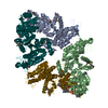


 PDBj
PDBj


 Assembly
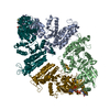


 Type: L-peptide linking / Mass: 147.129 Da / Num. of mol.: 2 / Source method: obtained synthetically / Formula: C5H9NO4
 Mass: 767.534 Da / Num. of mol.: 2 / Source method: obtained synthetically / Formula: C21H36N7O16P3S
 Mass: 18.015 Da / Num. of mol.: 6 / Source method: isolated from a natural source / Formula: H2O
 Sample preparation
 / Beamline: 22-ID / Wavelength: 1 Å
 Processing