 Movie
Movie Controller

[English] 日本語
 Yorodumi
- PDB-3s5c: Crystal Structure of a Hexachlorocyclohexane dehydrochlorinase (L... -
+ Open data

- Basic information
| Entry | Database: PDB / ID: 3s5c | ||||||
|---|---|---|---|---|---|---|---|
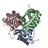
| Title | Crystal Structure of a Hexachlorocyclohexane dehydrochlorinase (LinA) Type2 | ||||||
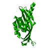
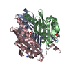
 Components Components | LinA | ||||||
 Keywords Keywords | TRANSFERASE / hexachlorocyclohexane dehydrochlorinase | ||||||
| Function / homology | SnoaL-like domain / SnoaL-like domain / Nuclear Transport Factor 2; Chain: A, - #50 / NTF2-like domain superfamily / Nuclear Transport Factor 2; Chain: A, / Roll / Alpha Beta / LinA Function and homology information Function and homology information | ||||||
| Biological species | uncultured organism (environmental samples) | ||||||
| Method |  X-RAY DIFFRACTION / MOLECULAR REPLACEMENT / Resolution: 3.5 Å X-RAY DIFFRACTION / MOLECULAR REPLACEMENT / Resolution: 3.5 Å | ||||||
 Authors Authors | Kukshal, V. / Macwan, A.S. / Kumar, A. / Ramachandran, R. | ||||||
 Citation Citation | Journal: Plos One / Year: 2012 Title: Crystal structure of the hexachlorocyclohexane dehydrochlorinase (LinA-type2): mutational analysis, thermostability and enantioselectivity Authors: Macwan, A.S. / Kukshal, V. / Srivastava, N. / Javed, S. / Kumar, A. / Ramachandran, R. | ||||||
| History |
|
- Structure visualization
| Structure viewer | Molecule: MolmilJmol/JSmol |
|---|
- Downloads & links
-Download
| PDBx/mmCIF format | 3s5c.cif.gz | 204.3 KB | Display | PDBx/mmCIF format |
|---|---|---|---|---|
| PDB format | pdb3s5c.ent.gz | 167.7 KB | Display | PDB format |
| PDBx/mmJSON format | 3s5c.json.gz | Tree view | PDBx/mmJSON format | |
| Others |  Other downloads Other downloads |
-Validation report
| Summary document | 3s5c_validation.pdf.gz | 466.8 KB | Display | wwPDB validaton report |
|---|---|---|---|---|
| Full document | 3s5c_full_validation.pdf.gz | 492.9 KB | Display | |
| Data in XML | 3s5c_validation.xml.gz | 37.6 KB | Display | |
| Data in CIF | 3s5c_validation.cif.gz | 51.5 KB | Display | |
| Arichive directory | https://data.pdbj.org/pub/pdb/validation_reports/s5/3s5cftp://data.pdbj.org/pub/pdb/validation_reports/s5/3s5c | HTTPS FTP |
-Related structure data
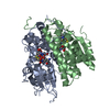
| Related structure data |  3a76S S: Starting model for refinement |
|---|---|
| Similar structure data |

-Links
 PDBj
PDBj
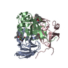
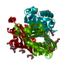
- Assembly
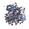
| Deposited unit | 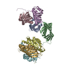
| ||||||||
|---|---|---|---|---|---|---|---|---|---|
| 1 | 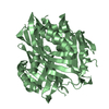
| ||||||||
| 2 | 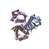
| ||||||||
| 3 | 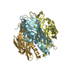
| ||||||||
| Unit cell |
|
-Components
| #1: Protein | Mass: 17335.467 Da / Num. of mol.: 7 Source method: isolated from a genetically manipulated source Details: The sequence was isolated from bacteria from a soil sample, metagenome source. It shows very high identity with the LinA from Sphingobium japonicum UT26 whose sequence is in PDB id: 3A76. Source: (gene. exp.) uncultured organism (environmental samples) Plasmid: pET-26b(+) / Production host:  |
|---|
-Experimental details
-Experiment
| Experiment | Method: X-RAY DIFFRACTION / Number of used crystals: 1 |
|---|
- Sample preparation
| Crystal | Density Matthews: 2.93 Å3/Da / Density % sol: 57.97 % |
|---|---|
| Crystal grow | Temperature: 295 K / Method: vapor diffusion, hanging drop / pH: 5.5 Details: 2% PEG 8000,0.1 M trisodium citrate, pH 5.5, VAPOR DIFFUSION, HANGING DROP, temperature 295K |
-Data collection
| Diffraction | Mean temperature: 295 K |
|---|---|
| Diffraction source | Source: ROTATING ANODE / Type: RIGAKU MICROMAX-007 HF / Wavelength: 1.5418 Å |
| Detector | Type: MAR scanner 345 mm plate / Detector: IMAGE PLATE / Date: Jun 24, 2010 |
| Radiation | Protocol: SINGLE WAVELENGTH / Monochromatic (M) / Laue (L): M / Scattering type: x-ray |
| Radiation wavelength | Wavelength: 1.5418 Å / Relative weight: 1 |
| Reflection | Resolution: 3.5→30.72 Å / Num. all: 18690 / Num. obs: 18690 / % possible obs: 99.1 % / Observed criterion σ(F): 8.7 / Observed criterion σ(I): 8.7 / Rmerge(I) obs: 0.239 |
| Reflection shell | Resolution: 3.5→3.69 Å / Redundancy: 4.6 % / Rmerge(I) obs: 0.663 / Mean I/σ(I) obs: 2.7 / Num. unique all: 2656 / % possible all: 98.5 |
- Processing
| Software |
| |||||||||||||||||||||||||||||||||||||||||||||||||||||||||||||||||
|---|---|---|---|---|---|---|---|---|---|---|---|---|---|---|---|---|---|---|---|---|---|---|---|---|---|---|---|---|---|---|---|---|---|---|---|---|---|---|---|---|---|---|---|---|---|---|---|---|---|---|---|---|---|---|---|---|---|---|---|---|---|---|---|---|---|---|
| Refinement | Method to determine structure: MOLECULAR REPLACEMENT Starting model: PDB ENTRY 3A76 Resolution: 3.5→30.72 Å / Cor.coef. Fo:Fc: 0.927 / Cor.coef. Fo:Fc free: 0.839 / Occupancy max: 1 / Occupancy min: 1 / SU B: 31.762 / SU ML: 0.486 / Cross valid method: THROUGHOUT / ESU R Free: 0.662 / Stereochemistry target values: MAXIMUM LIKELIHOOD Details: HYDROGENS HAVE BEEN ADDED IN THE RIDING POSITIONS U VALUES: REFINED INDIVIDUALLY
| |||||||||||||||||||||||||||||||||||||||||||||||||||||||||||||||||
| Solvent computation | Ion probe radii: 0.8 Å / Shrinkage radii: 0.8 Å / VDW probe radii: 1.4 Å / Solvent model: MASK | |||||||||||||||||||||||||||||||||||||||||||||||||||||||||||||||||
| Displacement parameters | Biso max: 105.35 Å2 / Biso mean: 50.1874 Å2 / Biso min: 18.29 Å2
| |||||||||||||||||||||||||||||||||||||||||||||||||||||||||||||||||
| Refinement step | Cycle: LAST / Resolution: 3.5→30.72 Å
| |||||||||||||||||||||||||||||||||||||||||||||||||||||||||||||||||
| Refine LS restraints |
| |||||||||||||||||||||||||||||||||||||||||||||||||||||||||||||||||
| LS refinement shell | Resolution: 3.5→3.59 Å / Total num. of bins used: 20
|
