Monochromator: double crystal / Protocol: SINGLE WAVELENGTH / Monochromatic (M) / Laue (L): M / Scattering type: x-ray
Radiation wavelength
Wavelength: 0.9791 Å / Relative weight: 1
Reflection
Resolution: 1.55→50 Å / Num. all: 22603 / Num. obs: 22441 / % possible obs: 99.3 % / Observed criterion σ(I): -3 / Redundancy: 7.5 % / Biso Wilson estimate: 18 Å2 / Rmerge(I) obs: 0.092 / Net I/σ(I): 24.3
Reflection shell
Resolution: 1.55→1.58 Å / Redundancy: 6.6 % / Rmerge(I) obs: 0.93 / Mean I/σ(I) obs: 2.1 / Num. unique all: 1110 / % possible all: 98.6
-
Processing
Software
Name
Version
Classification
SBC-Collect
datacollection
SHELX
modelbuilding
MLPHARE
phasing
DM
modelbuilding
ARP/wARP
modelbuilding
Coot
modelbuilding
PHENIX
(phenix.refine: dev_1096)
refinement
HKL-3000
datareduction
HKL-3000
datascaling
SHELX
phasing
DM
phasing
Refinement
Method to determine structure: SAD / Resolution: 1.55→28.974 Å / SU ML: 0.13 / Isotropic thermal model: isotropic / Cross valid method: Rfree / σ(F): 0 / Phase error: 16.09 / Stereochemistry target values: ML Details: HYDROGEN ATOMS HAVE BEEN ADDED AT THE RIDING POSITIONS
Rfactor
Num. reflection
% reflection
Selection details
Rfree
0.1798
1138
5.07 %
random
Rwork
0.1441
-
-
-
all
0.1458
22433
-
-
obs
0.1458
22433
99.37 %
-
Solvent computation
Shrinkage radii: 0.9 Å / VDW probe radii: 1.11 Å / Solvent model: FLAT BULK SOLVENT MODEL
Refinement step
Cycle: LAST / Resolution: 1.55→28.974 Å
Protein
Nucleic acid
Ligand
Solvent
Total
Num. atoms
1152
0
35
125
1312
Refine LS restraints
Refine-ID
Type
Dev ideal
Number
X-RAY DIFFRACTION
f_bond_d
0.013
1259
X-RAY DIFFRACTION
f_angle_d
1.391
1712
X-RAY DIFFRACTION
f_dihedral_angle_d
13.298
457
X-RAY DIFFRACTION
f_chiral_restr
0.086
185
X-RAY DIFFRACTION
f_plane_restr
0.007
224
LS refinement shell
Resolution (Å)
Rfactor Rfree
Num. reflection Rfree
Rfactor Rwork
Num. reflection Rwork
Refine-ID
% reflection obs (%)
1.5504-1.621
0.1946
136
0.1786
2646
X-RAY DIFFRACTION
99
1.621-1.7064
0.1743
158
0.1578
2626
X-RAY DIFFRACTION
99
1.7064-1.8133
0.2027
138
0.1456
2646
X-RAY DIFFRACTION
99
1.8133-1.9533
0.1661
144
0.1317
2650
X-RAY DIFFRACTION
99
1.9533-2.1498
0.1631
148
0.1205
2648
X-RAY DIFFRACTION
100
2.1498-2.4607
0.1681
146
0.1232
2665
X-RAY DIFFRACTION
100
2.4607-3.0997
0.1843
140
0.1423
2686
X-RAY DIFFRACTION
100
3.0997-28.979
0.1872
128
0.1574
2728
X-RAY DIFFRACTION
100
Refinement TLS params.
Method: refined / Refine-ID: X-RAY DIFFRACTION
ID
L11 (°2)
L12 (°2)
L13 (°2)
L22 (°2)
L23 (°2)
L33 (°2)
S11 (Å °)
S12 (Å °)
S13 (Å °)
S21 (Å °)
S22 (Å °)
S23 (Å °)
S31 (Å °)
S32 (Å °)
S33 (Å °)
T11 (Å2)
T12 (Å2)
T13 (Å2)
T22 (Å2)
T23 (Å2)
T33 (Å2)
Origin x (Å)
Origin y (Å)
Origin z (Å)
1
2.6564
0.2892
0.4828
3.0001
1.4981
0.7916
-0.0255
0.6115
0.1086
-0.8615
-0.0036
0.0193
-0.2806
0.0102
-0.128
0.1753
0.0026
0.0092
0.1519
-0.0149
0.0828
2.4317
26.9852
2.5831
2
0.2701
-0.1507
0.2075
0.1902
-0.2298
1.4506
0.0268
0.0201
-0.1715
-0.0291
-0.0053
-0.1272
0.1734
-0.0886
0.0931
0.0799
-0.0018
0.0006
0.0591
-0.0157
0.1106
-2.2273
17.0235
17.1668
3
2.3524
0.8143
1.3953
2.1508
1.5137
2.1836
0.0707
-0.2702
-0.3433
0.4458
0.0464
-0.2665
0.5812
-0.0575
0.1237
0.2224
0.0446
0.017
0.0808
0.0189
0.124
11.5895
14.0916
18.814
4
0.3221
-0.0589
0.1378
0.0593
0.1081
0.4233
-0.133
-0.1611
0.0134
0.1291
0.1101
-0.0508
0.0744
-0.0825
0.0232
0.2165
0.0695
0.0322
0.1025
-0.0133
0.1409
4.9183
9.3796
21.8485
5
1.4205
0.1238
0.0378
1.357
-0.1634
0.2049
-0.0007
-0.0671
-0.1708
-0.0446
0.0068
-0.0877
0.0731
0.0258
0.0268
0.0452
0
0.0042
0.0238
0.0051
0.0517
5.3496
23.1237
19.7447
6
3.2368
0.2479
-0.9452
0.932
0.6181
1.2321
-0.0734
-0.5864
-0.4653
0.2271
-0.0983
-0.0545
0.397
0.2393
-0.1605
0.1495
0.0217
-0.0175
0.1489
0.0441
0.1756
10.2789
23.7514
32.2943
Refinement TLS group
ID
Refine-ID
Refine TLS-ID
Selection details
1
X-RAY DIFFRACTION
1
(chainAandresid2:12)
2
X-RAY DIFFRACTION
2
(chainAandresid13:30)
3
X-RAY DIFFRACTION
3
(chainAandresid31:51)
4
X-RAY DIFFRACTION
4
(chainAandresid52:67)
5
X-RAY DIFFRACTION
5
(chainAandresid68:134)
6
X-RAY DIFFRACTION
6
(chainAandresid135:149)
+
About Yorodumi
-
News
-
Feb 9, 2022. New format data for meta-information of EMDB entries
New format data for meta-information of EMDB entries
Version 3 of the EMDB header file is now the official format.
The previous official version 1.9 will be removed from the archive.
In the structure databanks used in Yorodumi, some data are registered as the other names, "COVID-19 virus" and "2019-nCoV". Here are the details of the virus and the list of structure data.
Jan 31, 2019. EMDB accession codes are about to change! (news from PDBe EMDB page)
EMDB accession codes are about to change! (news from PDBe EMDB page)
The allocation of 4 digits for EMDB accession codes will soon come to an end. Whilst these codes will remain in use, new EMDB accession codes will include an additional digit and will expand incrementally as the available range of codes is exhausted. The current 4-digit format prefixed with “EMD-” (i.e. EMD-XXXX) will advance to a 5-digit format (i.e. EMD-XXXXX), and so on. It is currently estimated that the 4-digit codes will be depleted around Spring 2019, at which point the 5-digit format will come into force.
The EM Navigator/Yorodumi systems omit the EMD- prefix.
Related info.:Q: What is EMD? / ID/Accession-code notation in Yorodumi/EM Navigator
Yorodumi is a browser for structure data from EMDB, PDB, SASBDB, etc.
This page is also the successor to EM Navigator detail page, and also detail information page/front-end page for Omokage search.
The word "yorodu" (or yorozu) is an old Japanese word meaning "ten thousand". "mi" (miru) is to see.
Related info.:EMDB / PDB / SASBDB / Comparison of 3 databanks / Yorodumi Search / Aug 31, 2016. New EM Navigator & Yorodumi / Yorodumi Papers / Jmol/JSmol / Function and homology information / Changes in new EM Navigator and Yorodumi
 Movie
Movie Controller


 Open data
 Basic information
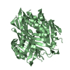
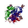
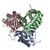
 Components
 Keywords
 Function and homology information
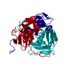
 Kribbella flavida (bacteria)
 X-RAY DIFFRACTION /
  Authors
 Citation
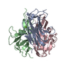
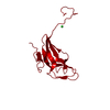
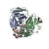
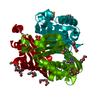
 Structure visualization
 Downloads & links
 Other downloads
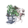
 PDBj
PDBj
 Assembly

 Mass: 94.971 Da / Num. of mol.: 1 / Source method: obtained synthetically / Formula: PO4
 Mass: 150.173 Da / Num. of mol.: 3 / Source method: obtained synthetically / Formula: C6H14O4
 Mass: 18.015 Da / Num. of mol.: 125 / Source method: isolated from a natural source / Formula: H2O
 Sample preparation
 / Beamline: 19-BM / Wavelength: 0.9791 Å
 Processing