 Movie
Movie Controller

[English] 日本語
 Yorodumi
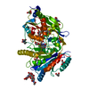
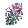
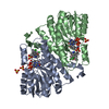
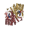
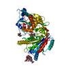
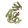
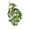
- PDB-3s1c: Maize cytokinin oxidase/dehydrogenase complexed with N6-isopenten... -
+ Open data

- Basic information
| Entry | Database: PDB / ID: 3s1c | |||||||||
|---|---|---|---|---|---|---|---|---|---|---|
| Title | Maize cytokinin oxidase/dehydrogenase complexed with N6-isopentenyladenosine | |||||||||
 Components Components | Cytokinin dehydrogenase 1 | |||||||||
 Keywords Keywords | OXIDOREDUCTASE / FAD BINDING PROTEIN / FLAVOPROTEIN / CYTOKININ OXIDASE/DEHYDROGENASE / CYTOKININ BINDING / GLYCOSYLATION / COVALENT FLAVINATION | |||||||||
| Function / homology |  Function and homology information Function and homology informationcytokinin dehydrogenase / cytokinin dehydrogenase activity / cytokinin metabolic process / FAD binding / oxidoreductase activity / extracellular region Similarity search - Function | |||||||||
| Biological species |  | |||||||||
| Method |  X-RAY DIFFRACTION / SYNCHROTRON / MOLECULAR REPLACEMENT / Resolution: 2.09 Å X-RAY DIFFRACTION / SYNCHROTRON / MOLECULAR REPLACEMENT / Resolution: 2.09 Å | |||||||||
 Authors Authors | Kopecny, D. / Briozzo, P. / Morera, S. | |||||||||
 Citation Citation | Journal: Febs J. / Year: 2016 Title: Kinetic and structural investigation of the cytokinin oxidase/dehydrogenase active site. Authors: Kopecny, D. / Koncitikova, R. / Popelka, H. / Briozzo, P. / Vigouroux, A. / Kopecna, M. / Zalabak, D. / Sebela, M. / Skopalova, J. / Frebort, I. / Morera, S. | |||||||||
| History |
|
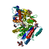
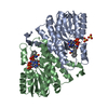
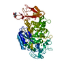
- Structure visualization
| Structure viewer | Molecule: MolmilJmol/JSmol |
|---|
- Downloads & links
-Download
| PDBx/mmCIF format | 3s1c.cif.gz | 134.1 KB | Display | PDBx/mmCIF format |
|---|---|---|---|---|
| PDB format | pdb3s1c.ent.gz | 94.7 KB | Display | PDB format |
| PDBx/mmJSON format | 3s1c.json.gz | Tree view | PDBx/mmJSON format | |
| Others |  Other downloads Other downloads |
-Validation report
| Arichive directory | https://data.pdbj.org/pub/pdb/validation_reports/s1/3s1cftp://data.pdbj.org/pub/pdb/validation_reports/s1/3s1c | HTTPS FTP |
|---|
-Related structure data
| Related structure data |  3s1dC  3s1eC 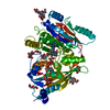 3s1fC 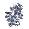 4ml8C 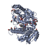 4mlaC 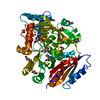 4o95C  4oalC  3bw7S S: Starting model for refinement C: citing same article ( |
|---|---|
| Similar structure data |


-Links
 PDBj
PDBj
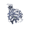
- Assembly
| Deposited unit | 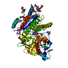
| ||||||||
|---|---|---|---|---|---|---|---|---|---|
| 1 |
| ||||||||
| Unit cell |
|
-Components
-Protein , 1 types, 1 molecules A
| #1: Protein | Mass: 55357.184 Da / Num. of mol.: 1 Source method: isolated from a genetically manipulated source Source: (gene. exp.)  Yarrowia lipolytica (yeast) / Strain (production host): Po1g / References: UniProt: Q9T0N8, cytokinin dehydrogenase Yarrowia lipolytica (yeast) / Strain (production host): Po1g / References: UniProt: Q9T0N8, cytokinin dehydrogenase |
|---|
-Sugars , 2 types, 3 molecules 
| #2: Polysaccharide | 2-acetamido-2-deoxy-beta-D-glucopyranose-(1-4)-2-acetamido-2-deoxy-beta-D-glucopyranose Source method: isolated from a genetically manipulated source |
|---|---|
| #8: Sugar |  Type: D-saccharide, beta linking / Mass: 221.208 Da / Num. of mol.: 2 Type: D-saccharide, beta linking / Mass: 221.208 Da / Num. of mol.: 2Source method: isolated from a genetically manipulated source Formula: C8H15NO6 |
-Non-polymers , 6 types, 348 molecules 


| #3: Chemical | ChemComp-FAD /  Mass: 785.550 Da / Num. of mol.: 1 / Source method: obtained synthetically / Formula: C27H33N9O15P2 / Comment: FAD*YM Mass: 785.550 Da / Num. of mol.: 1 / Source method: obtained synthetically / Formula: C27H33N9O15P2 / Comment: FAD*YM | ||||||
|---|---|---|---|---|---|---|---|
| #4: Chemical | ChemComp-ZIR /  Mass: 335.358 Da / Num. of mol.: 1 / Source method: obtained synthetically / Formula: C15H21N5O4 Mass: 335.358 Da / Num. of mol.: 1 / Source method: obtained synthetically / Formula: C15H21N5O4 | ||||||
| #5: Chemical | ChemComp-GOL /  Mass: 92.094 Da / Num. of mol.: 14 / Source method: obtained synthetically / Formula: C3H8O3 Mass: 92.094 Da / Num. of mol.: 14 / Source method: obtained synthetically / Formula: C3H8O3#6: Chemical | ChemComp-15P /  Mass: 1529.829 Da / Num. of mol.: 4 / Source method: obtained synthetically / Formula: C69H140O35 / Comment: precipitant*YM Mass: 1529.829 Da / Num. of mol.: 4 / Source method: obtained synthetically / Formula: C69H140O35 / Comment: precipitant*YM#7: Chemical | ChemComp-PEG /  Mass: 106.120 Da / Num. of mol.: 4 / Source method: obtained synthetically / Formula: C4H10O3 Mass: 106.120 Da / Num. of mol.: 4 / Source method: obtained synthetically / Formula: C4H10O3#9: Water | ChemComp-HOH / | Mass: 18.015 Da / Num. of mol.: 324 / Source method: isolated from a natural source / Formula: H2O |
-Details
| Has protein modification | Y |
|---|---|
| Sequence details | SEQUENCE CONFLICT IN UNP ENTRY Q9T0N8 AT THESE POSITIONS |
-Experimental details
-Experiment
| Experiment | Method: X-RAY DIFFRACTION / Number of used crystals: 1 |
|---|
- Sample preparation
| Crystal | Density Matthews: 2.94 Å3/Da / Density % sol: 58.19 % |
|---|---|
| Crystal grow | Temperature: 293 K / Method: vapor diffusion, hanging drop / pH: 7 Details: 30% (w/v) PEG 1500, 0.5% (w/v)n-octyl beta-D-glucoside, 0.1 M Tris-HCl, pH 7.0, VAPOR DIFFUSION, HANGING DROP, temperature 293.0K |
-Data collection
| Diffraction | Mean temperature: 100 K | ||||||||||||||||||||||||||||||||||||||||||||||||||||||||||||||||||
|---|---|---|---|---|---|---|---|---|---|---|---|---|---|---|---|---|---|---|---|---|---|---|---|---|---|---|---|---|---|---|---|---|---|---|---|---|---|---|---|---|---|---|---|---|---|---|---|---|---|---|---|---|---|---|---|---|---|---|---|---|---|---|---|---|---|---|---|
| Diffraction source | Source: SYNCHROTRON / Site: ESRF  / Beamline: BM30A / Wavelength: 0.979629 Å / Beamline: BM30A / Wavelength: 0.979629 Å | ||||||||||||||||||||||||||||||||||||||||||||||||||||||||||||||||||
| Detector | Type: ADSC QUANTUM 315 / Detector: CCD / Date: Jul 2, 2005 | ||||||||||||||||||||||||||||||||||||||||||||||||||||||||||||||||||
| Radiation | Monochromator: not known / Protocol: SINGLE WAVELENGTH / Monochromatic (M) / Laue (L): M / Scattering type: x-ray | ||||||||||||||||||||||||||||||||||||||||||||||||||||||||||||||||||
| Radiation wavelength | Wavelength: 0.979629 Å / Relative weight: 1 | ||||||||||||||||||||||||||||||||||||||||||||||||||||||||||||||||||
| Reflection | Resolution: 2.09→35 Å / Num. all: 38547 / Num. obs: 35133 / % possible obs: 91.1 % / Observed criterion σ(F): 2 / Observed criterion σ(I): 1 / Biso Wilson estimate: 35.04 Å2 / Rmerge(I) obs: 0.058 / Net I/σ(I): 16.9 | ||||||||||||||||||||||||||||||||||||||||||||||||||||||||||||||||||
| Reflection shell |
|
- Processing
| Software |
| ||||||||||||||||||||||||||||||||||||||||||||||||||||||||||||||||||||||||
|---|---|---|---|---|---|---|---|---|---|---|---|---|---|---|---|---|---|---|---|---|---|---|---|---|---|---|---|---|---|---|---|---|---|---|---|---|---|---|---|---|---|---|---|---|---|---|---|---|---|---|---|---|---|---|---|---|---|---|---|---|---|---|---|---|---|---|---|---|---|---|---|---|---|
| Refinement | Method to determine structure: MOLECULAR REPLACEMENT Starting model: PDB entry 3BW7 Resolution: 2.09→32.5 Å / Cor.coef. Fo:Fc: 0.9132 / Cor.coef. Fo:Fc free: 0.8643 / Cross valid method: THROUGHOUT / σ(F): 0 / Stereochemistry target values: Engh & Huber
| ||||||||||||||||||||||||||||||||||||||||||||||||||||||||||||||||||||||||
| Displacement parameters | Biso mean: 30.63 Å2
| ||||||||||||||||||||||||||||||||||||||||||||||||||||||||||||||||||||||||
| Refine analyze | Luzzati coordinate error obs: 0.286 Å | ||||||||||||||||||||||||||||||||||||||||||||||||||||||||||||||||||||||||
| Refinement step | Cycle: LAST / Resolution: 2.09→32.5 Å
| ||||||||||||||||||||||||||||||||||||||||||||||||||||||||||||||||||||||||
| Refine LS restraints |
| ||||||||||||||||||||||||||||||||||||||||||||||||||||||||||||||||||||||||
| LS refinement shell | Resolution: 2.09→2.15 Å / Total num. of bins used: 18
|
