 Movie
Movie Controller


+ Open data

- Basic information
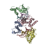
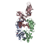
| Entry | Database: PDB / ID: 3ry6 | |||||||||
|---|---|---|---|---|---|---|---|---|---|---|
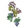
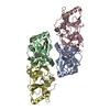
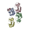
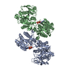
| Title | Complex of fcgammariia (CD32) and the FC of human IGG1 | |||||||||
 Components Components |
| |||||||||
 Keywords Keywords | IMMUNE SYSTEM / FC RECEPTOR / CD32 / IMMUNOGLOBULIN SUPERFAMILY / HIGH RESPONDER POLYMORPHISM / HUMAN IGG1 / THERAPEUTIC ANTIBODY / GLYCOPROTEIN / IMMUNOGLOBULIN C REGION / IMMUNOGLOBULIN DOMAIN / CELL MEMBRANE / IGG-BINDING PROTEIN / MEMBRANE / PHOSPHOPROTEIN / RECEPTOR / TRANSMEMBRANE | |||||||||
| Function / homology |  Function and homology information Function and homology informationIgG receptor activity / complement-dependent cytotoxicity / antibody-dependent cellular cytotoxicity / Fc-gamma receptor I complex binding / IgG binding / immunoglobulin complex, circulating / Classical antibody-mediated complement activation / immunoglobulin receptor binding / IgG immunoglobulin complex / Initial triggering of complement ...IgG receptor activity / complement-dependent cytotoxicity / antibody-dependent cellular cytotoxicity / Fc-gamma receptor I complex binding / IgG binding / immunoglobulin complex, circulating / Classical antibody-mediated complement activation / immunoglobulin receptor binding / IgG immunoglobulin complex / Initial triggering of complement / FCGR activation / complement activation, classical pathway / Role of phospholipids in phagocytosis / antigen binding / FCGR3A-mediated IL10 synthesis / positive regulation of phagocytosis / Regulation of Complement cascade / secretory granule membrane / B cell receptor signaling pathway / FCGR3A-mediated phagocytosis / Regulation of actin dynamics for phagocytic cup formation / positive regulation of tumor necrosis factor production / antibacterial humoral response / Interleukin-4 and Interleukin-13 signaling / blood microparticle / adaptive immune response / cell surface receptor signaling pathway / external side of plasma membrane / Neutrophil degranulation / : / extracellular exosome / extracellular region / plasma membrane Similarity search - Function | |||||||||
| Biological species |  Homo sapiens (human) Homo sapiens (human) | |||||||||
| Method |  X-RAY DIFFRACTION / MOLECULAR REPLACEMENT / Resolution: 3.8 Å X-RAY DIFFRACTION / MOLECULAR REPLACEMENT / Resolution: 3.8 Å | |||||||||
 Authors Authors | Ramsland, P.A. / Farrugia, W. / Scott, A.M. / Hogarth, P.M. | |||||||||
 Citation Citation | Journal: J.Immunol. / Year: 2011 Title: Structural Basis for Fc{gamma}RIIa Recognition of Human IgG and Formation of Inflammatory Signaling Complexes. Authors: Ramsland, P.A. / Farrugia, W. / Bradford, T.M. / Sardjono, C.T. / Esparon, S. / Trist, H.M. / Powell, M.S. / Tan, P.S. / Cendron, A.C. / Wines, B.D. / Scott, A.M. / Hogarth, P.M. | |||||||||
| History |
|
- Structure visualization
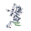
| Structure viewer | Molecule: MolmilJmol/JSmol |
|---|
- Downloads & links
-Download
| PDBx/mmCIF format | 3ry6.cif.gz | 138.3 KB | Display | PDBx/mmCIF format |
|---|---|---|---|---|
| PDB format | pdb3ry6.ent.gz | 107.4 KB | Display | PDB format |
| PDBx/mmJSON format | 3ry6.json.gz | Tree view | PDBx/mmJSON format | |
| Others |  Other downloads Other downloads |
-Validation report
| Arichive directory | https://data.pdbj.org/pub/pdb/validation_reports/ry/3ry6ftp://data.pdbj.org/pub/pdb/validation_reports/ry/3ry6 | HTTPS FTP |
|---|
-Related structure data
| Related structure data |  3ry4C  3ry5C  1e4kS C: citing same article ( S: Starting model for refinement |
|---|---|
| Similar structure data |

-Links
 PDBj
PDBj


- Assembly
| Deposited unit | 
| ||||||||
|---|---|---|---|---|---|---|---|---|---|
| 1 |
| ||||||||
| Unit cell |
|
-Components
-Protein , 2 types, 3 molecules ABC
| #1: Protein | Mass: 24244.457 Da / Num. of mol.: 2 / Fragment: UNP residues 114-327 Source method: isolated from a genetically manipulated source Source: (gene. exp.) Homo sapiens (human) / Gene: IGHG1 / Cell line (production host): NS0 MURINE MYELOMA CELLS / Production host:  #2: Protein | | Mass: 18881.088 Da / Num. of mol.: 1 / Fragment: UNP residues 40-206 Source method: isolated from a genetically manipulated source Source: (gene. exp.) Homo sapiens (human) / Gene: CD32, FCG2, FCGR2A, FCGR2A1, IGFR2 / Plasmid: PVL1392 / Cell line (production host): SF21 / Production host:   Spodoptera frugiperda (fall armyworm) / References: UniProt: P12318 Spodoptera frugiperda (fall armyworm) / References: UniProt: P12318 |
|---|
-Sugars , 4 types, 4 molecules 
| #3: Polysaccharide | N-acetyl-alpha-neuraminic acid-(2-6)-beta-D-galactopyranose-(1-4)-2-acetamido-2-deoxy-beta-D- ...N-acetyl-alpha-neuraminic acid-(2-6)-beta-D-galactopyranose-(1-4)-2-acetamido-2-deoxy-beta-D-glucopyranose-(1-2)-alpha-D-mannopyranose-(1-3)-[2-acetamido-2-deoxy-beta-D-glucopyranose-(1-2)-alpha-D-mannopyranose-(1-6)]beta-D-mannopyranose-(1-4)-2-acetamido-2-deoxy-beta-D-glucopyranose-(1-4)-[beta-L-fucopyranose-(1-6)]2-acetamido-2-deoxy-alpha-D-glucopyranose Source method: isolated from a genetically manipulated source |
|---|---|
| #4: Polysaccharide | N-acetyl-alpha-neuraminic acid-(2-6)-beta-D-galactopyranose-(1-4)-2-acetamido-2-deoxy-beta-D- ...N-acetyl-alpha-neuraminic acid-(2-6)-beta-D-galactopyranose-(1-4)-2-acetamido-2-deoxy-beta-D-glucopyranose-(1-2)-alpha-D-mannopyranose-(1-3)-[N-acetyl-alpha-neuraminic acid-(2-6)-beta-D-galactopyranose-(1-4)-2-acetamido-2-deoxy-beta-D-glucopyranose-(1-2)-alpha-D-mannopyranose-(1-6)]beta-D-mannopyranose-(1-4)-2-acetamido-2-deoxy-beta-D-glucopyranose-(1-4)-[beta-L-fucopyranose-(1-6)]2-acetamido-2-deoxy-beta-D-glucopyranose Source method: isolated from a genetically manipulated source |
| #5: Polysaccharide | beta-D-mannopyranose-(1-3)-[alpha-D-mannopyranose-(1-6)]alpha-D-mannopyranose-(1-4)-2-acetamido-2- ...beta-D-mannopyranose-(1-3)-[alpha-D-mannopyranose-(1-6)]alpha-D-mannopyranose-(1-4)-2-acetamido-2-deoxy-beta-D-glucopyranose-(1-4)-[beta-L-fucopyranose-(1-6)]2-acetamido-2-deoxy-alpha-D-glucopyranose Source method: isolated from a genetically manipulated source |
| #7: Sugar | ChemComp-NAG /  Type: D-saccharide, beta linking / Mass: 221.208 Da / Num. of mol.: 1 Type: D-saccharide, beta linking / Mass: 221.208 Da / Num. of mol.: 1Source method: isolated from a genetically manipulated source Formula: C8H15NO6 |
-Non-polymers , 1 types, 4 molecules 
| #6: Chemical | ChemComp-GOL /  Mass: 92.094 Da / Num. of mol.: 4 / Source method: obtained synthetically / Formula: C3H8O3 Mass: 92.094 Da / Num. of mol.: 4 / Source method: obtained synthetically / Formula: C3H8O3 |
|---|
-Details
| Has protein modification | Y |
|---|---|
| Sequence details | THERE ARE VARIANT D239E (VAR_002887), VARIANT L241M (VAR_003888) IN G1M(NON-1) MARKER. EU DIFFERS ...THERE ARE VARIANT D239E (VAR_002887), VARIANT L241M (VAR_003888) IN G1M(NON-1) MARKER. EU DIFFERS IN THE AMIDATION STATES OF RESIDUES 155, 166, 177, 195, 198 IN REFERENCE: BIOCHEMIST |
-Experimental details
-Experiment
| Experiment | Method: X-RAY DIFFRACTION / Number of used crystals: 1 |
|---|
- Sample preparation
| Crystal | Density Matthews: 4.25 Å3/Da / Density % sol: 71.04 % |
|---|---|
| Crystal grow | Method: vapor diffusion / pH: 7.4 Details: 20% (W/V) PEG 3350, 5MM TRIS, 0.15M NACL, 2MM MOPS, 0.2M TRI-POTASSIUM ACETATE, PH 7.40, VAPOR DIFFUSION, TEMPERATURE 291.0K |
-Data collection
| Diffraction | Mean temperature: 100 K |
|---|---|
| Diffraction source | Source: ROTATING ANODE / Type: RIGAKU / Wavelength: 1.5418 |
| Detector | Type: RIGAKU RAXIS IV / Detector: IMAGE PLATE / Date: Apr 1, 2004 / Details: MIRRORS |
| Radiation | Monochromator: OSMIC BLUE MIRRORS / Protocol: SINGLE WAVELENGTH / Monochromatic (M) / Laue (L): M / Scattering type: x-ray |
| Radiation wavelength | Wavelength: 1.5418 Å / Relative weight: 1 |
| Reflection | Resolution: 3.8→30 Å / Num. obs: 11766 / % possible obs: 98.7 % / Observed criterion σ(I): 0 / Redundancy: 5 % / Biso Wilson estimate: 134.6 Å2 / Rsym value: 0.137 / Net I/σ(I): 10.8 |
| Reflection shell | Resolution: 3.8→3.94 Å / Redundancy: 4.8 % / Mean I/σ(I) obs: 3.78 / Rsym value: 0.35 / % possible all: 99.7 |
- Processing
| Software |
| ||||||||||||||||||||||||||||||||||||||||||||||||||||||||||||
|---|---|---|---|---|---|---|---|---|---|---|---|---|---|---|---|---|---|---|---|---|---|---|---|---|---|---|---|---|---|---|---|---|---|---|---|---|---|---|---|---|---|---|---|---|---|---|---|---|---|---|---|---|---|---|---|---|---|---|---|---|---|
| Refinement | Method to determine structure: MOLECULAR REPLACEMENT Starting model: PDB ENTRY 1E4K Resolution: 3.8→29.71 Å / Cross valid method: THROUGHOUT / σ(F): 0 / Stereochemistry target values: ENGH & HUBER
| ||||||||||||||||||||||||||||||||||||||||||||||||||||||||||||
| Refine analyze |
| ||||||||||||||||||||||||||||||||||||||||||||||||||||||||||||
| Refinement step | Cycle: LAST / Resolution: 3.8→29.71 Å
| ||||||||||||||||||||||||||||||||||||||||||||||||||||||||||||
| Refine LS restraints |
| ||||||||||||||||||||||||||||||||||||||||||||||||||||||||||||
| LS refinement shell | Resolution: 3.8→3.94 Å / Rfactor Rfree error: 0.055 /
|
