 Movie
Movie Controller

[English] 日本語
 Yorodumi
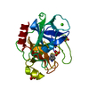
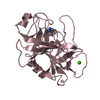
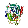
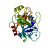
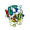
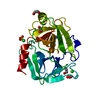
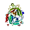
- PDB-3rxd: Crystal structure of Trypsin complexed with (3-methoxyphenyl)meth... -
+ Open data

- Basic information
| Entry | Database: PDB / ID: 3rxd | ||||||
|---|---|---|---|---|---|---|---|
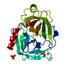
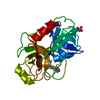
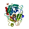
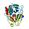
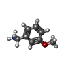
| Title | Crystal structure of Trypsin complexed with (3-methoxyphenyl)methanamine | ||||||
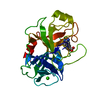
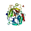
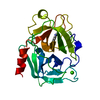
 Components Components | Cationic trypsin | ||||||
 Keywords Keywords | HYDROLASE/HYDROLASE INHIBITOR / Trypsin-like serine proteases / HYDROLASE-HYDROLASE INHIBITOR complex | ||||||
| Function / homology |  Function and homology information Function and homology informationtrypsin / serpin family protein binding / serine protease inhibitor complex / digestion / endopeptidase activity / serine-type endopeptidase activity / proteolysis / : / metal ion binding Similarity search - Function | ||||||
| Biological species |  | ||||||
| Method |  X-RAY DIFFRACTION / MOLECULAR REPLACEMENT / Resolution: 1.7 Å X-RAY DIFFRACTION / MOLECULAR REPLACEMENT / Resolution: 1.7 Å | ||||||
 Authors Authors | Yamane, J. / Yao, M. / Zhou, Y. / Tanaka, I. | ||||||
 Citation Citation | Journal: J.Appl.Crystallogr. / Year: 2011 Title: In-crystal affinity ranking of fragment hit compounds reveals a relationship with their inhibitory activities Authors: Yamane, J. / Yao, M. / Zhou, Y. / Hiramatsu, Y. / Fujiwara, K. / Yamaguchi, T. / Yamaguchi, H. / Togame, H. / Tsujishita, H. / Takemoto, H. / Tanaka, I. | ||||||
| History |
|
- Structure visualization
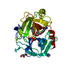
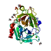
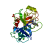
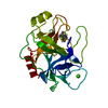
| Structure viewer | Molecule: MolmilJmol/JSmol |
|---|
- Downloads & links
-Download
| PDBx/mmCIF format | 3rxd.cif.gz | 63.5 KB | Display | PDBx/mmCIF format |
|---|---|---|---|---|
| PDB format | pdb3rxd.ent.gz | 44.3 KB | Display | PDB format |
| PDBx/mmJSON format | 3rxd.json.gz | Tree view | PDBx/mmJSON format | |
| Others |  Other downloads Other downloads |
-Validation report
| Arichive directory | https://data.pdbj.org/pub/pdb/validation_reports/rx/3rxdftp://data.pdbj.org/pub/pdb/validation_reports/rx/3rxd | HTTPS FTP |
|---|
-Related structure data
| Related structure data | 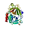 3atiC 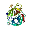 3atkC  3atlC 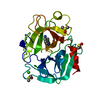 3atmC 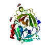 3rxaC 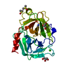 3rxbC 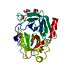 3rxcC 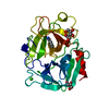 3rxeC 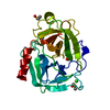 3rxfC 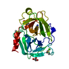 3rxgC 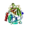 3rxhC  3rxiC 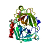 3rxjC 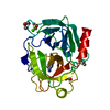 3rxkC  3rxlC 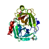 3rxmC 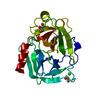 3rxoC 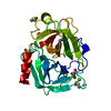 3rxpC 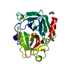 3rxqC 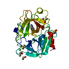 3rxrC 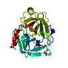 3rxsC 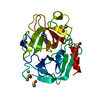 3rxtC  3rxuC 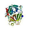 3rxvC 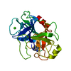 1s0rS C: citing same article ( S: Starting model for refinement |
|---|---|
| Similar structure data |


-Links
 PDBj
PDBj

- Assembly
| Deposited unit | 
| ||||||||
|---|---|---|---|---|---|---|---|---|---|
| 1 |
| ||||||||
| Unit cell |
|
-Components
-Protein , 1 types, 1 molecules A
| #1: Protein | Mass: 23324.287 Da / Num. of mol.: 1 / Source method: isolated from a natural source / Source: (natural) |
|---|
-Non-polymers , 5 types, 316 molecules 


| #2: Chemical | ChemComp-CA /  Mass: 40.078 Da / Num. of mol.: 1 / Source method: obtained synthetically / Formula: Ca Mass: 40.078 Da / Num. of mol.: 1 / Source method: obtained synthetically / Formula: Ca | ||||||
|---|---|---|---|---|---|---|---|
| #3: Chemical |  Mass: 92.094 Da / Num. of mol.: 2 / Source method: obtained synthetically / Formula: C3H8O3 Mass: 92.094 Da / Num. of mol.: 2 / Source method: obtained synthetically / Formula: C3H8O3#4: Chemical |  Mass: 78.133 Da / Num. of mol.: 2 / Source method: obtained synthetically / Formula: C2H6OS / Comment: DMSO, precipitant*YM Mass: 78.133 Da / Num. of mol.: 2 / Source method: obtained synthetically / Formula: C2H6OS / Comment: DMSO, precipitant*YM#5: Chemical | ChemComp-SZ4 / |  Mass: 137.179 Da / Num. of mol.: 1 / Source method: obtained synthetically / Formula: C8H11NO Mass: 137.179 Da / Num. of mol.: 1 / Source method: obtained synthetically / Formula: C8H11NO#6: Water | ChemComp-HOH / | Mass: 18.015 Da / Num. of mol.: 310 / Source method: isolated from a natural source / Formula: H2O |
-Details
| Has protein modification | Y |
|---|
-Experimental details
-Experiment
| Experiment | Method: X-RAY DIFFRACTION / Number of used crystals: 1 |
|---|
- Sample preparation
| Crystal | Density Matthews: 2.26 Å3/Da / Density % sol: 45.63 % |
|---|---|
| Crystal grow | Temperature: 277 K / Method: vapor diffusion, sitting drop / pH: 8.5 Details: 0.1M Tris-HCl, 30% PEG 3350, 0.2M Lithium Sulfate, pH 8.5, VAPOR DIFFUSION, SITTING DROP, temperature 277K |
-Data collection
| Diffraction | Mean temperature: 100 K |
|---|---|
| Diffraction source | Source: ROTATING ANODE / Type: RIGAKU MICROMAX-007 / Wavelength: 1.5418 Å |
| Detector | Type: RIGAKU RAXIS IV++ / Detector: IMAGE PLATE |
| Radiation | Protocol: SINGLE WAVELENGTH / Monochromatic (M) / Laue (L): M / Scattering type: x-ray |
| Radiation wavelength | Wavelength: 1.5418 Å / Relative weight: 1 |
| Reflection | Resolution: 1.7→50 Å / Num. all: 23997 / Num. obs: 23517 / % possible obs: 98 % / Observed criterion σ(I): -3 / Redundancy: 4.7 % / Biso Wilson estimate: 16.5 Å2 / Rmerge(I) obs: 0.044 / Rsym value: 0.044 |
- Processing
| Software |
| ||||||||||||||||||||||||||||||||||||||||||||||||||||||||||||
|---|---|---|---|---|---|---|---|---|---|---|---|---|---|---|---|---|---|---|---|---|---|---|---|---|---|---|---|---|---|---|---|---|---|---|---|---|---|---|---|---|---|---|---|---|---|---|---|---|---|---|---|---|---|---|---|---|---|---|---|---|---|
| Refinement | Method to determine structure: MOLECULAR REPLACEMENT Starting model: PDB ENTRY 1S0R Resolution: 1.7→20 Å / Cor.coef. Fo:Fc: 0.96 / Cor.coef. Fo:Fc free: 0.954 / SU B: 1.682 / SU ML: 0.057 / Cross valid method: THROUGHOUT / ESU R Free: 0.099 / Stereochemistry target values: MAXIMUM LIKELIHOOD / Details: HYDROGENS HAVE BEEN USED IF PRESENT IN THE INPUT
| ||||||||||||||||||||||||||||||||||||||||||||||||||||||||||||
| Solvent computation | Ion probe radii: 0.8 Å / Shrinkage radii: 0.8 Å / VDW probe radii: 1.2 Å / Solvent model: BABINET MODEL WITH MASK | ||||||||||||||||||||||||||||||||||||||||||||||||||||||||||||
| Displacement parameters | Biso mean: 13.044 Å2
| ||||||||||||||||||||||||||||||||||||||||||||||||||||||||||||
| Refinement step | Cycle: LAST / Resolution: 1.7→20 Å
| ||||||||||||||||||||||||||||||||||||||||||||||||||||||||||||
| Refine LS restraints |
| ||||||||||||||||||||||||||||||||||||||||||||||||||||||||||||
| LS refinement shell | Resolution: 1.7→1.744 Å / Rfactor Rfree error: 0.099 / Total num. of bins used: 20
|
