 Movie
Movie Controller


+ Open data

- Basic information
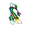
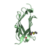
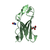
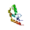
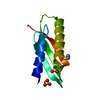
| Entry | Database: PDB / ID: 3rp1 | ||||||
|---|---|---|---|---|---|---|---|
| Title | Crystal structure of Human LAIR-1 in C2 space group | ||||||
 Components Components | Leukocyte-associated immunoglobulin-like receptor 1 | ||||||
 Keywords Keywords | IMMUNE SYSTEM / structural genomics / collagen receptor / New York Structural Genomics Research Consortium / NYSGRC / PSI-Biology / Ig-like / collagen-binding | ||||||
| Function / homology |  Function and homology information Function and homology informationimmune response-regulating signaling pathway / tertiary granule membrane / specific granule membrane / Immunoregulatory interactions between a Lymphoid and a non-Lymphoid cell / adaptive immune response / Neutrophil degranulation / plasma membrane Similarity search - Function | ||||||
| Biological species |  Homo sapiens (human) Homo sapiens (human) | ||||||
| Method |  X-RAY DIFFRACTION / SYNCHROTRON / MOLECULAR REPLACEMENT / Resolution: 2.6 Å X-RAY DIFFRACTION / SYNCHROTRON / MOLECULAR REPLACEMENT / Resolution: 2.6 Å | ||||||
 Authors Authors | Sampathkumar, P. / Ramagopal, U.A. / Yan, Q. / Toro, R. / Nathenson, S. / Bonanno, J. / Almo, S.C. / New York Structural Genomics Research Consortium (NYSGRC) | ||||||
 Citation Citation | Journal: To be Published Title: Crystal structure of Human LAIR-1 in C2 space group Authors: Sampathkumar, P. / Ramagopal, U.A. / Yan, Q. / Toro, R. / Nathenson, S. / Bonanno, J. / Almo, S.C. | ||||||
| History |
|
- Structure visualization
| Structure viewer | Molecule: MolmilJmol/JSmol |
|---|
- Downloads & links
-Download
| PDBx/mmCIF format | 3rp1.cif.gz | 162 KB | Display | PDBx/mmCIF format |
|---|---|---|---|---|
| PDB format | pdb3rp1.ent.gz | 129.4 KB | Display | PDB format |
| PDBx/mmJSON format | 3rp1.json.gz | Tree view | PDBx/mmJSON format | |
| Others |  Other downloads Other downloads |
-Validation report
| Arichive directory | https://data.pdbj.org/pub/pdb/validation_reports/rp/3rp1ftp://data.pdbj.org/pub/pdb/validation_reports/rp/3rp1 | HTTPS FTP |
|---|
-Related structure data
| Related structure data |  3kgrS S: Starting model for refinement |
|---|---|
| Similar structure data | |
| Other databases |


-Links
 PDBj
PDBj
- Assembly
| Deposited unit | 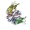
| ||||||||
|---|---|---|---|---|---|---|---|---|---|
| 1 | 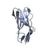
| ||||||||
| 2 | 
| ||||||||
| 3 | 
| ||||||||
| 4 | 
| ||||||||
| Unit cell |
|
-Components
| #1: Protein | Mass: 11611.771 Da / Num. of mol.: 4 / Fragment: Extracellular domain (UNP residues 22-123) Source method: isolated from a genetically manipulated source Source: (gene. exp.) Homo sapiens (human) / Gene: CD305, LAIR-1, LAIR1 / Plasmid: pET3a / Production host:  #2: Water | ChemComp-HOH / |  Mass: 18.015 Da / Num. of mol.: 33 / Source method: isolated from a natural source / Formula: H2O Mass: 18.015 Da / Num. of mol.: 33 / Source method: isolated from a natural source / Formula: H2OHas protein modification | Y | |
|---|
-Experimental details
-Experiment
| Experiment | Method: X-RAY DIFFRACTION / Number of used crystals: 1 |
|---|
- Sample preparation
| Crystal | Density Matthews: 2.13 Å3/Da / Density % sol: 42.18 % |
|---|---|
| Crystal grow | Temperature: 298 K / Method: vapor diffusion, sitting drop / pH: 7.5 Details: 200mM calcium choloride, 28% PEG400, 100mM HEPES, pH 7.5, VAPOR DIFFUSION, SITTING DROP, temperature 298K |
-Data collection
| Diffraction | Mean temperature: 100 K |
|---|---|
| Diffraction source | Source: SYNCHROTRON / Site: NSLS  / Beamline: X29A / Wavelength: 1.081 Å / Beamline: X29A / Wavelength: 1.081 Å |
| Detector | Type: ADSC QUANTUM 315 / Detector: CCD / Date: Aug 19, 2010 |
| Radiation | Protocol: SINGLE WAVELENGTH / Monochromatic (M) / Laue (L): M / Scattering type: x-ray |
| Radiation wavelength | Wavelength: 1.081 Å / Relative weight: 1 |
| Reflection | Resolution: 2.6→50 Å / Num. obs: 12005 / % possible obs: 99.9 % / Redundancy: 7.2 % / Biso Wilson estimate: 59.1 Å2 / Rsym value: 0.112 / Net I/σ(I): 17.8 |
| Reflection shell | Resolution: 2.6→2.69 Å / Redundancy: 7.3 % / Mean I/σ(I) obs: 4 / Num. unique all: 1173 / Rsym value: 0.639 / % possible all: 99.9 |
- Processing
| Software |
| |||||||||||||||||||||||||||||||||||||||||||||||||||||||||||||||||||||||||||||||||||||||||||||||||||||||||||||||||||||||||||||
|---|---|---|---|---|---|---|---|---|---|---|---|---|---|---|---|---|---|---|---|---|---|---|---|---|---|---|---|---|---|---|---|---|---|---|---|---|---|---|---|---|---|---|---|---|---|---|---|---|---|---|---|---|---|---|---|---|---|---|---|---|---|---|---|---|---|---|---|---|---|---|---|---|---|---|---|---|---|---|---|---|---|---|---|---|---|---|---|---|---|---|---|---|---|---|---|---|---|---|---|---|---|---|---|---|---|---|---|---|---|---|---|---|---|---|---|---|---|---|---|---|---|---|---|---|---|---|
| Refinement | Method to determine structure: MOLECULAR REPLACEMENT Starting model: 3KGR Resolution: 2.6→31.82 Å / Cor.coef. Fo:Fc: 0.932 / Cor.coef. Fo:Fc free: 0.866 / Occupancy max: 1 / Occupancy min: 0.5 / SU B: 32.562 / SU ML: 0.317 / Cross valid method: THROUGHOUT / σ(F): 0 / ESU R Free: 0.41 / Stereochemistry target values: MAXIMUM LIKELIHOOD Details: HYDROGENS HAVE BEEN ADDED IN THE RIDING POSITIONS. U VALUES: WITH TLS ADDED
| |||||||||||||||||||||||||||||||||||||||||||||||||||||||||||||||||||||||||||||||||||||||||||||||||||||||||||||||||||||||||||||
| Solvent computation | Ion probe radii: 0.8 Å / Shrinkage radii: 0.8 Å / VDW probe radii: 1.4 Å / Solvent model: BABINET MODEL WITH MASK | |||||||||||||||||||||||||||||||||||||||||||||||||||||||||||||||||||||||||||||||||||||||||||||||||||||||||||||||||||||||||||||
| Displacement parameters | Biso max: 92.98 Å2 / Biso mean: 42.1602 Å2 / Biso min: 18.94 Å2
| |||||||||||||||||||||||||||||||||||||||||||||||||||||||||||||||||||||||||||||||||||||||||||||||||||||||||||||||||||||||||||||
| Refinement step | Cycle: LAST / Resolution: 2.6→31.82 Å
| |||||||||||||||||||||||||||||||||||||||||||||||||||||||||||||||||||||||||||||||||||||||||||||||||||||||||||||||||||||||||||||
| Refine LS restraints |
| |||||||||||||||||||||||||||||||||||||||||||||||||||||||||||||||||||||||||||||||||||||||||||||||||||||||||||||||||||||||||||||
| LS refinement shell | Resolution: 2.602→2.669 Å / Total num. of bins used: 20
| |||||||||||||||||||||||||||||||||||||||||||||||||||||||||||||||||||||||||||||||||||||||||||||||||||||||||||||||||||||||||||||
| Refinement TLS params. | Method: refined / Refine-ID: X-RAY DIFFRACTION
| |||||||||||||||||||||||||||||||||||||||||||||||||||||||||||||||||||||||||||||||||||||||||||||||||||||||||||||||||||||||||||||
| Refinement TLS group |
|
