Mass: 18.015 Da / Num. of mol.: 78 / Source method: isolated from a natural source / Formula: H2O
Has protein modification
Y
-
Experimental details
-
Experiment
Experiment
Method: X-RAY DIFFRACTION / Number of used crystals: 1
-
Sample preparation
Crystal
Density Matthews: 3.25 Å3/Da / Density % sol: 62.19 %
Crystal grow
Temperature: 297 K / Method: vapor diffusion, hanging drop / pH: 8 Details: 1.8 M NaCl, 0.1 M Tris/HCl, VAPOR DIFFUSION, HANGING DROP, temperature 297K
Monochromator: double crystal / Protocol: SINGLE WAVELENGTH / Monochromatic (M) / Laue (L): M / Scattering type: x-ray
Radiation wavelength
Wavelength: 0.97923 Å / Relative weight: 1
Reflection
Resolution: 1.85→50 Å / Num. all: 14603 / Num. obs: 14408 / % possible obs: 98.7 % / Observed criterion σ(I): -3 / Redundancy: 5 % / Biso Wilson estimate: 22 Å2 / Rmerge(I) obs: 0.095 / Net I/σ(I): 25.6
Reflection shell
Resolution: 1.85→1.88 Å / Redundancy: 4.5 % / Rmerge(I) obs: 0.646 / Mean I/σ(I) obs: 2.6 / Num. unique all: 707 / % possible all: 95.9
-
Processing
Software
Name
Version
Classification
SBC-Collect
datacollection
SHELX
modelbuilding
MLPHARE
phasing
DM
modelbuilding
ARP/wARP
modelbuilding
Coot
modelbuilding
PHENIX
(phenix.refine: 1.6.4_486)
refinement
HKL-3000
datareduction
HKL-3000
datascaling
SHELX
phasing
DM
phasing
Refinement
Method to determine structure: SAD / Resolution: 1.85→25.932 Å / SU ML: 0.18 / Isotropic thermal model: isotropic / Cross valid method: THROUGHOUT / σ(F): 1.36 / Stereochemistry target values: ML Details: Hydrogen atoms have been added at ridding positions
Rfactor
Num. reflection
% reflection
Selection details
Rfree
0.1908
1102
7.65 %
random
Rwork
0.1617
-
-
-
all
0.1639
14402
-
-
obs
0.1639
14402
98.8 %
-
Solvent computation
Shrinkage radii: 0.53 Å / VDW probe radii: 0.7 Å / Solvent model: FLAT BULK SOLVENT MODEL / Bsol: 50.816 Å2 / ksol: 0.482 e/Å3
Displacement parameters
Baniso -1
Baniso -2
Baniso -3
1-
-0.0213 Å2
-0 Å2
0 Å2
2-
-
-0.0213 Å2
-0 Å2
3-
-
-
0.0426 Å2
Refinement step
Cycle: LAST / Resolution: 1.85→25.932 Å
Protein
Nucleic acid
Ligand
Solvent
Total
Num. atoms
876
0
16
78
970
Refine LS restraints
Refine-ID
Type
Dev ideal
Number
X-RAY DIFFRACTION
f_bond_d
0.015
941
X-RAY DIFFRACTION
f_angle_d
1.381
1282
X-RAY DIFFRACTION
f_dihedral_angle_d
14.957
343
X-RAY DIFFRACTION
f_chiral_restr
0.1
136
X-RAY DIFFRACTION
f_plane_restr
0.007
166
LS refinement shell
Refine-ID: X-RAY DIFFRACTION
Resolution (Å)
Rfactor Rfree
Num. reflection Rfree
Rfactor Rwork
Num. reflection Rwork
Num. reflection obs
% reflection obs (%)
1.8472-1.9313
0.2481
142
0.2143
1644
1644
98
1.9313-2.0331
0.2211
132
0.1751
1623
1623
98
2.0331-2.1604
0.1979
151
0.1579
1632
1632
99
2.1604-2.3271
0.1873
129
0.1557
1675
1675
99
2.3271-2.5611
0.2041
142
0.1611
1656
1656
99
2.5611-2.9313
0.2225
135
0.1862
1669
1669
99
2.9313-3.6914
0.188
139
0.1695
1673
1673
100
3.6914-25.935
0.1526
132
0.1359
1728
1728
99
Refinement TLS params.
Method: refined / Refine-ID: X-RAY DIFFRACTION
ID
L11 (°2)
L12 (°2)
L13 (°2)
L22 (°2)
L23 (°2)
L33 (°2)
S11 (Å °)
S12 (Å °)
S13 (Å °)
S21 (Å °)
S22 (Å °)
S23 (Å °)
S31 (Å °)
S32 (Å °)
S33 (Å °)
T11 (Å2)
T12 (Å2)
T13 (Å2)
T22 (Å2)
T23 (Å2)
T33 (Å2)
Origin x (Å)
Origin y (Å)
Origin z (Å)
1
1.132
0.6553
0.1689
0.656
-0.0911
0.1629
0.0772
0.0232
0.0171
0.1016
-0.0646
-0.0336
-0.0422
0.0468
-0.0055
0.1394
-0.0259
-0.014
0.1469
-0.0019
0.1319
4.2905
-34.0524
6.4667
2
0.5337
0.9601
-0.196
2.1653
0.0384
0.5018
0.2219
-0.1429
0.0967
0.4127
-0.2266
0.1306
-0.0274
0.06
0.0099
0.2685
-0.0551
-0.0083
0.175
-0.0221
0.1791
0.0861
-30.7172
12.6263
3
0.9737
0.4367
-0.1492
1.4248
-0.1879
0.1271
0.0363
-0.02
0.0192
0.0522
-0.0227
0.1638
0.0065
0.1066
0.0007
0.1176
0.0099
-0.0091
0.1481
-0.007
0.1075
-3.4849
-39.7442
8.8042
4
4.0479
-0.206
0.4253
2.5183
0.5433
0.1731
-0.0195
1.5556
0.1487
-0.8975
-0.1433
-0.0402
-0.3617
0.0968
0.1004
0.3642
0.0416
-0.0401
0.5147
-0.0015
0.1628
-2.977
-32.1733
-11.3643
5
2.6469
-0.4737
-0.2309
2.5167
-1.0767
2.0326
0.3772
0.4406
-0.2317
0.1695
-0.3317
-0.6202
-0.2645
-0.427
-0.0796
0.161
0.0767
-0.0138
0.1991
0.032
0.2645
-10.6832
-28.4979
-1.6035
6
3.6653
-0.8987
-0.3387
0.6872
-0.3289
0.4138
0.6882
1.0842
0.2594
-0.5075
-0.486
-0.1906
0.0165
0.0897
-0.1185
0.3393
0.1649
0.0817
0.4258
0.1151
0.3076
-12.1633
-24.3925
-9.0422
7
2.0933
2.3291
4.4376
3.0439
5.0093
9.4769
-0.3479
0.3841
-0.3494
0.195
0.03
0.1037
0.0099
0.4171
0.2088
0.2436
0.064
0.0337
0.3324
-0.0537
0.2011
-19.9386
-31.0991
-9.5811
Refinement TLS group
ID
Refine-ID
Refine TLS-ID
Selection details
1
X-RAY DIFFRACTION
1
(chainAandresid1:23)
2
X-RAY DIFFRACTION
2
(chainAandresid24:39)
3
X-RAY DIFFRACTION
3
(chainAandresid40:64)
4
X-RAY DIFFRACTION
4
(chainAandresid65:78)
5
X-RAY DIFFRACTION
5
(chainAandresid79:96)
6
X-RAY DIFFRACTION
6
(chainAandresid97:107)
7
X-RAY DIFFRACTION
7
(chainAandresid108:111)
+
About Yorodumi
-
News
-
Feb 9, 2022. New format data for meta-information of EMDB entries
New format data for meta-information of EMDB entries
Version 3 of the EMDB header file is now the official format.
The previous official version 1.9 will be removed from the archive.
In the structure databanks used in Yorodumi, some data are registered as the other names, "COVID-19 virus" and "2019-nCoV". Here are the details of the virus and the list of structure data.
Jan 31, 2019. EMDB accession codes are about to change! (news from PDBe EMDB page)
EMDB accession codes are about to change! (news from PDBe EMDB page)
The allocation of 4 digits for EMDB accession codes will soon come to an end. Whilst these codes will remain in use, new EMDB accession codes will include an additional digit and will expand incrementally as the available range of codes is exhausted. The current 4-digit format prefixed with “EMD-” (i.e. EMD-XXXX) will advance to a 5-digit format (i.e. EMD-XXXXX), and so on. It is currently estimated that the 4-digit codes will be depleted around Spring 2019, at which point the 5-digit format will come into force.
The EM Navigator/Yorodumi systems omit the EMD- prefix.
Related info.:Q: What is EMD? / ID/Accession-code notation in Yorodumi/EM Navigator
Yorodumi is a browser for structure data from EMDB, PDB, SASBDB, etc.
This page is also the successor to EM Navigator detail page, and also detail information page/front-end page for Omokage search.
The word "yorodu" (or yorozu) is an old Japanese word meaning "ten thousand". "mi" (miru) is to see.
Related info.:EMDB / PDB / SASBDB / Comparison of 3 databanks / Yorodumi Search / Aug 31, 2016. New EM Navigator & Yorodumi / Yorodumi Papers / Jmol/JSmol / Function and homology information / Changes in new EM Navigator and Yorodumi
 Movie
Movie Controller

 Yorodumi
 Open data
 Basic information
 Components
 Keywords
 Function and homology information
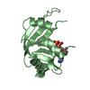
 Saccharomonospora viridis (bacteria)
 X-RAY DIFFRACTION /
  Authors
 Citation
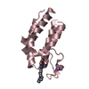
 Structure visualization
 Downloads & links
 Other downloads
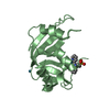


 PDBj
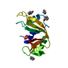
PDBj Assembly

 Mass: 65.409 Da / Num. of mol.: 1 / Source method: obtained synthetically / Formula: Zn
 Mass: 122.143 Da / Num. of mol.: 1 / Source method: obtained synthetically / Formula: C4H12NO3 / Comment: pH buffer*YM
 Mass: 35.453 Da / Num. of mol.: 7 / Source method: obtained synthetically / Formula: Cl
 Mass: 18.015 Da / Num. of mol.: 78 / Source method: isolated from a natural source / Formula: H2O
 Sample preparation
 / Beamline: 19-BM / Wavelength: 0.97923 Å
 Processing