 Movie
Movie Controller


+ Open data

- Basic information
| Entry | Database: PDB / ID: 3rgo | ||||||
|---|---|---|---|---|---|---|---|
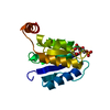
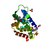
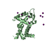
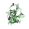
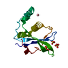
| Title | Crystal Structure of PTPMT1 | ||||||
 Components Components | Protein-tyrosine phosphatase mitochondrial 1 | ||||||
 Keywords Keywords | HYDROLASE / phosphatidylglycerol phosphate (PGP) phosphatase | ||||||
| Function / homology |  Function and homology information Function and homology informationphosphatidylinositol-5-phosphate phosphatase activity / phosphatidylglycerophosphatase / phosphatidylglycerophosphatase activity / cardiolipin biosynthetic process / regulation of intrinsic apoptotic signaling pathway / phosphatidylinositol-4,5-bisphosphate 5-phosphatase activity / mitochondrial fragmentation involved in apoptotic process / phosphatidylinositol metabolic process / negative regulation of insulin secretion involved in cellular response to glucose stimulus / protein-serine/threonine phosphatase ...phosphatidylinositol-5-phosphate phosphatase activity / phosphatidylglycerophosphatase / phosphatidylglycerophosphatase activity / cardiolipin biosynthetic process / regulation of intrinsic apoptotic signaling pathway / phosphatidylinositol-4,5-bisphosphate 5-phosphatase activity / mitochondrial fragmentation involved in apoptotic process / phosphatidylinositol metabolic process / negative regulation of insulin secretion involved in cellular response to glucose stimulus / protein-serine/threonine phosphatase / protein serine/threonine phosphatase activity / protein-tyrosine-phosphatase / protein tyrosine phosphatase activity / mitochondrial inner membrane / mitochondrion Similarity search - Function | ||||||
| Biological species |  | ||||||
| Method |  X-RAY DIFFRACTION / SYNCHROTRON / MAD / Resolution: 1.928 Å X-RAY DIFFRACTION / SYNCHROTRON / MAD / Resolution: 1.928 Å | ||||||
 Authors Authors | Xiao, J. / Engel, J.L. | ||||||
 Citation Citation | Journal: Proc.Natl.Acad.Sci.USA / Year: 2011 Title: Structural and functional analysis of PTPMT1, a phosphatase required for cardiolipin synthesis. Authors: Xiao, J. / Engel, J.L. / Zhang, J. / Chen, M.J. / Manning, G. / Dixon, J.E. | ||||||
| History |
|
- Structure visualization
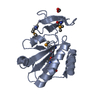
| Structure viewer | Molecule: MolmilJmol/JSmol |
|---|
- Downloads & links
-Download
| PDBx/mmCIF format | 3rgo.cif.gz | 78.3 KB | Display | PDBx/mmCIF format |
|---|---|---|---|---|
| PDB format | pdb3rgo.ent.gz | 58.2 KB | Display | PDB format |
| PDBx/mmJSON format | 3rgo.json.gz | Tree view | PDBx/mmJSON format | |
| Others |  Other downloads Other downloads |
-Validation report
| Arichive directory | https://data.pdbj.org/pub/pdb/validation_reports/rg/3rgoftp://data.pdbj.org/pub/pdb/validation_reports/rg/3rgo | HTTPS FTP |
|---|
-Related structure data


-Links
 PDBj
PDBj


- Assembly
| Deposited unit | 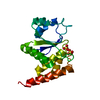
| ||||||||
|---|---|---|---|---|---|---|---|---|---|
| 1 |
| ||||||||
| Unit cell |
|
-Components
| #1: Protein | Mass: 17926.816 Da / Num. of mol.: 1 Source method: isolated from a genetically manipulated source Source: (gene. exp.)  References: UniProt: Q66GT5, protein-serine/threonine phosphatase, protein-tyrosine-phosphatase | ||
|---|---|---|---|
| #2: Chemical |   Mass: 96.063 Da / Num. of mol.: 2 / Source method: obtained synthetically / Formula: SO4 Mass: 96.063 Da / Num. of mol.: 2 / Source method: obtained synthetically / Formula: SO4#3: Water | ChemComp-HOH / |  Mass: 18.015 Da / Num. of mol.: 48 / Source method: isolated from a natural source / Formula: H2O Mass: 18.015 Da / Num. of mol.: 48 / Source method: isolated from a natural source / Formula: H2O |
-Experimental details
-Experiment
| Experiment | Method: X-RAY DIFFRACTION / Number of used crystals: 1 |
|---|
- Sample preparation
| Crystal | Density Matthews: 2.13 Å3/Da / Density % sol: 42.29 % |
|---|---|
| Crystal grow | Temperature: 277 K / Method: vapor diffusion, sitting drop / pH: 5.5 Details: PEG 5000 MME, Bis-Tris, (NH4)2SO4, pH 5.5, VAPOR DIFFUSION, SITTING DROP, temperature 277K |
-Data collection
| Diffraction |
| |||||||||||||||||||||||||||||||||||||||||||||||||||||||||||||||||||||||||||||||||||||||||||||||||||||||||||||||||||||||||||||||||||||||||||||||||||
|---|---|---|---|---|---|---|---|---|---|---|---|---|---|---|---|---|---|---|---|---|---|---|---|---|---|---|---|---|---|---|---|---|---|---|---|---|---|---|---|---|---|---|---|---|---|---|---|---|---|---|---|---|---|---|---|---|---|---|---|---|---|---|---|---|---|---|---|---|---|---|---|---|---|---|---|---|---|---|---|---|---|---|---|---|---|---|---|---|---|---|---|---|---|---|---|---|---|---|---|---|---|---|---|---|---|---|---|---|---|---|---|---|---|---|---|---|---|---|---|---|---|---|---|---|---|---|---|---|---|---|---|---|---|---|---|---|---|---|---|---|---|---|---|---|---|---|---|---|
| Diffraction source |
| |||||||||||||||||||||||||||||||||||||||||||||||||||||||||||||||||||||||||||||||||||||||||||||||||||||||||||||||||||||||||||||||||||||||||||||||||||
| Detector |
| |||||||||||||||||||||||||||||||||||||||||||||||||||||||||||||||||||||||||||||||||||||||||||||||||||||||||||||||||||||||||||||||||||||||||||||||||||
| Radiation |
| |||||||||||||||||||||||||||||||||||||||||||||||||||||||||||||||||||||||||||||||||||||||||||||||||||||||||||||||||||||||||||||||||||||||||||||||||||
| Radiation wavelength |
| |||||||||||||||||||||||||||||||||||||||||||||||||||||||||||||||||||||||||||||||||||||||||||||||||||||||||||||||||||||||||||||||||||||||||||||||||||
| Reflection | Redundancy: 7.4 % / Av σ(I) over netI: 61.88 / Number: 86345 / Rmerge(I) obs: 0.063 / Χ2: 1.83 / D res high: 2 Å / D res low: 50 Å / Num. obs: 11679 / % possible obs: 99.9 | |||||||||||||||||||||||||||||||||||||||||||||||||||||||||||||||||||||||||||||||||||||||||||||||||||||||||||||||||||||||||||||||||||||||||||||||||||
| Diffraction reflection shell |
| |||||||||||||||||||||||||||||||||||||||||||||||||||||||||||||||||||||||||||||||||||||||||||||||||||||||||||||||||||||||||||||||||||||||||||||||||||
| Reflection | Resolution: 1.928→50 Å / Num. obs: 12762 / % possible obs: 99.9 % / Redundancy: 12.7 % / Rmerge(I) obs: 0.051 / Χ2: 1.224 / Net I/σ(I): 12.1 | |||||||||||||||||||||||||||||||||||||||||||||||||||||||||||||||||||||||||||||||||||||||||||||||||||||||||||||||||||||||||||||||||||||||||||||||||||
| Reflection shell |
|

-Phasing
| Phasing | Method: MAD |
|---|
- Processing
| Software |
| ||||||||||||||||||||||||||||||||||||||||
|---|---|---|---|---|---|---|---|---|---|---|---|---|---|---|---|---|---|---|---|---|---|---|---|---|---|---|---|---|---|---|---|---|---|---|---|---|---|---|---|---|---|
| Refinement | Method to determine structure: MAD / Resolution: 1.928→34.749 Å / Occupancy max: 1 / Occupancy min: 1 / FOM work R set: 0.8212 / SU ML: 0.25 / σ(F): 1.34 / Stereochemistry target values: ML
| ||||||||||||||||||||||||||||||||||||||||
| Solvent computation | Shrinkage radii: 0.9 Å / VDW probe radii: 1.11 Å / Solvent model: FLAT BULK SOLVENT MODEL / Bsol: 46.546 Å2 / ksol: 0.34 e/Å3 | ||||||||||||||||||||||||||||||||||||||||
| Displacement parameters | Biso max: 186.99 Å2 / Biso mean: 51.7747 Å2 / Biso min: 30.38 Å2
| ||||||||||||||||||||||||||||||||||||||||
| Refinement step | Cycle: LAST / Resolution: 1.928→34.749 Å
| ||||||||||||||||||||||||||||||||||||||||
| Refine LS restraints |
| ||||||||||||||||||||||||||||||||||||||||
| LS refinement shell | Refine-ID: X-RAY DIFFRACTION / Total num. of bins used: 4
| ||||||||||||||||||||||||||||||||||||||||
| Refinement TLS params. | Method: refined / Origin x: 13.1083 Å / Origin y: -1.3176 Å / Origin z: -15.3095 Å
| ||||||||||||||||||||||||||||||||||||||||
| Refinement TLS group | Selection details: ALL |
