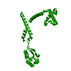
- PDB-3rfw: The virulence factor PEB4 and the periplasmic protein Cj1289 are ... -
+
Open data
ID or keywords:
Loading...
-
Basic information
Entry
Database: PDB / ID: 3rfw
Title
The virulence factor PEB4 and the periplasmic protein Cj1289 are two structurally-related SurA-like chaperones in the human pathogen Campylobacter jejuni
Mass: 18.015 Da / Num. of mol.: 144 / Source method: isolated from a natural source / Formula: H2O
-
Experimental details
-
Experiment
Experiment
Method: X-RAY DIFFRACTION / Number of used crystals: 2
-
Sample preparation
Crystal
Density Matthews: 3.94 Å3/Da / Density % sol: 68.8 % / Mosaicity: 0 °
Crystal grow
Temperature: 280 K / Method: vapor diffusion, sitting drop / pH: 3.5 Details: 0.1M succinate, 40% v/v Jeffamine, pH 3.5, VAPOR DIFFUSION, SITTING DROP, temperature 280K
-
Data collection
Diffraction
ID
Mean temperature (K)
Crystal-ID
1
100
1
2
100
1
Diffraction source
Source
Site
Beamline
ID
Wavelength (Å)
SYNCHROTRON
Diamond
I02
1
0.978
2
0.9800, 0.9803, 0.9745
Detector
Type: ADSC QUANTUM 315 / Detector: CCD
Radiation
ID
Protocol
Monochromatic (M) / Laue (L)
Scattering type
Wavelength-ID
1
SINGLEWAVELENGTH
M
x-ray
1
2
MAD
M
x-ray
1
Radiation wavelength
ID
Wavelength (Å)
Relative weight
1
0.978
1
2
0.98
1
3
0.9803
1
4
0.9745
1
Reflection
Redundancy: 20.4 % / Av σ(I) over netI: 3.8 / Number: 277591 / Rsym value: 0.084 / D res high: 2.6 Å / D res low: 59.996 Å / Num. obs: 13585 / % possible obs: 100
Diffraction reflection shell
Highest resolution (Å)
Lowest resolution (Å)
% possible obs (%)
ID
Rmerge(I) obs
Rsym value
Redundancy
8.22
45.9
99.4
1
0.078
0.078
19.9
5.81
8.22
100
1
0.053
0.053
21.3
4.75
5.81
100
1
0.056
0.056
21.2
4.11
4.75
100
1
0.056
0.056
21.5
3.68
4.11
100
1
0.074
0.074
21.4
3.36
3.68
100
1
0.094
0.094
21.5
3.11
3.36
100
1
0.116
0.116
21.7
2.91
3.11
100
1
0.166
0.166
21.7
2.74
2.91
100
1
0.291
0.291
20.9
2.6
2.74
100
1
0.507
0.507
15.2
Reflection
Resolution: 2.199→45.797 Å / Num. all: 22275 / Num. obs: 22275 / % possible obs: 99.5 % / Redundancy: 11.3 % / Biso Wilson estimate: 41.7 Å2 / Rsym value: 0.07 / Net I/σ(I): 20.3
Reflection shell
Resolution (Å)
Redundancy (%)
Rmerge(I) all
Rmerge(I) obs
Mean I/σ(I) obs
Num. measured all
Num. unique all
Rpim(I) all
Rrim(I) all
Rsym value
Net I/σ(I) obs
% possible all
2.2-2.32
11.4
0.464
0.443
1.4
36924
3238
0.138
0.464
0.443
5.7
100
2.32-2.46
11.6
0.254
0.242
3
35926
3099
0.074
0.254
0.242
7.9
100
2.46-2.63
11.6
0.18
0.172
4.1
33369
2881
0.053
0.18
0.172
10.9
100
2.63-2.84
11.5
0.13
0.124
5.4
30885
2693
0.039
0.13
0.124
15.4
100
2.84-3.11
11.4
0.082
0.079
7.8
28365
2487
0.025
0.082
0.079
22.2
100
3.11-3.48
11.4
0.065
0.062
9.5
25663
2252
0.019
0.065
0.062
29.4
100
3.48-4.02
10.9
0.064
0.061
9.6
21836
2005
0.019
0.064
0.061
35.7
100
4.02-4.92
10.2
0.059
0.056
10.4
17067
1670
0.019
0.059
0.056
38.7
100
4.92-6.96
11.8
0.054
0.051
10.9
15620
1324
0.016
0.054
0.051
41.4
100
6.96-45.797
8.8
0.055
0.051
11.8
5478
626
0.02
0.055
0.051
36.8
85.4
-
Phasing
Phasing
Method: MAD
Phasing set
ID
1
2
3
Phasing MAD
D res high: 2.69 Å / D res low: 20 Å / FOM : 0.55 / Reflection: 12185
Phasing MAD set
Clust-ID
Expt-ID
Set-ID
Wavelength (Å)
F double prime refined
F prime refined
3wavelength
1
1
0.98
3.5
-7.8
3wavelength
1
2
0.9803
4.96
-7.11
3wavelength
1
3
0.9745
3.5
-5
Phasing MAD set site
ID
Atom type symbol
B iso
Fract x
Fract y
Fract z
Occupancy
1
Se
60
0.024
0
0.156
1.446
2
Se
60
0.563
0.041
0.265
1.143
3
Se
60
0.883
0.086
0.253
0.977
Phasing MAD shell
Resolution (Å)
FOM
Reflection
9.07-20
0.8
644
5.94-9.07
0.83
1028
4.71-5.94
0.74
1280
4.02-4.71
0.69
1505
3.56-4.02
0.59
1681
3.23-3.56
0.49
1868
2.98-3.23
0.39
2023
2.78-2.98
0.29
2156
-
Processing
Software
Name
Version
Classification
NB
SCALA
3.3.9
datascaling
SOLVE
2.13
phasing
REFMAC
refinement
PDB_EXTRACT
3.1
dataextraction
Refinement
Method to determine structure: MAD / Resolution: 2.2→39.75 Å / Cor.coef. Fo:Fc: 0.936 / Cor.coef. Fo:Fc free: 0.913 / WRfactor Rfree: 0.289 / WRfactor Rwork: 0.2495 / Occupancy max: 1 / Occupancy min: 1 / FOM work R set: 0.7905 / SU B: 5.556 / SU ML: 0.143 / SU R Cruickshank DPI: 0.209 / SU Rfree: 0.1931 / Cross valid method: THROUGHOUT / ESU R Free: 0.193 / Stereochemistry target values: MAXIMUM LIKELIHOOD Details: HYDROGENS HAVE BEEN ADDED IN THE RIDING POSITIONS U VALUES: REFINED INDIVIDUALLY
Rfactor
Num. reflection
% reflection
Selection details
Rfree
0.2763
1139
5.1 %
RANDOM
Rwork
0.2341
-
-
-
obs
0.2362
21134
99.24 %
-
Solvent computation
Ion probe radii: 0.8 Å / Shrinkage radii: 0.8 Å / VDW probe radii: 1.4 Å / Solvent model: MASK
In the structure databanks used in Yorodumi, some data are registered as the other names, "COVID-19 virus" and "2019-nCoV". Here are the details of the virus and the list of structure data.
Jan 31, 2019. EMDB accession codes are about to change! (news from PDBe EMDB page)
EMDB accession codes are about to change! (news from PDBe EMDB page)
The allocation of 4 digits for EMDB accession codes will soon come to an end. Whilst these codes will remain in use, new EMDB accession codes will include an additional digit and will expand incrementally as the available range of codes is exhausted. The current 4-digit format prefixed with “EMD-” (i.e. EMD-XXXX) will advance to a 5-digit format (i.e. EMD-XXXXX), and so on. It is currently estimated that the 4-digit codes will be depleted around Spring 2019, at which point the 5-digit format will come into force.
The EM Navigator/Yorodumi systems omit the EMD- prefix.
Related info.:Q: What is EMD? / ID/Accession-code notation in Yorodumi/EM Navigator
Yorodumi is a browser for structure data from EMDB, PDB, SASBDB, etc.
This page is also the successor to EM Navigator detail page, and also detail information page/front-end page for Omokage search.
The word "yorodu" (or yorozu) is an old Japanese word meaning "ten thousand". "mi" (miru) is to see.
Related info.:EMDB / PDB / SASBDB / Comparison of 3 databanks / Yorodumi Search / Aug 31, 2016. New EM Navigator & Yorodumi / Yorodumi Papers / Jmol/JSmol / Function and homology information / Changes in new EM Navigator and Yorodumi
 Movie
Movie Controller

 Yorodumi
 Open data
 Basic information
 Components
 Keywords
 Function and homology information
 Campylobacter jejuni (Campylobacter)
 X-RAY DIFFRACTION /
  Authors
 Citation
 Structure visualization
 Downloads & links
 Other downloads
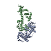


 PDBj
PDBj


 Assembly
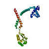
 Mass: 18.015 Da / Num. of mol.: 144 / Source method: isolated from a natural source / Formula: H2O
 Sample preparation
 Processing