 Movie
Movie Controller

[English] 日本語
 Yorodumi
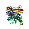
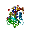
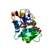
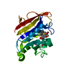
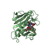
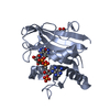
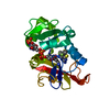
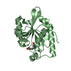
- PDB-3rbi: The Type III Crystal Structure of Streptococcus agalactiae Sortase C1 -
+ Open data

- Basic information
| Entry | Database: PDB / ID: 3rbi | ||||||
|---|---|---|---|---|---|---|---|
| Title | The Type III Crystal Structure of Streptococcus agalactiae Sortase C1 | ||||||
 Components Components | Sortase family protein | ||||||
 Keywords Keywords | HYDROLASE / Sortase fold / Beta-barrel / Class C sortase / Pili biogenesis | ||||||
| Function / homology |  Function and homology information Function and homology information | ||||||
| Biological species |  Streptococcus agalactiae serogroup V (bacteria) Streptococcus agalactiae serogroup V (bacteria) | ||||||
| Method |  X-RAY DIFFRACTION / SYNCHROTRON / MOLECULAR REPLACEMENT / Resolution: 3 Å X-RAY DIFFRACTION / SYNCHROTRON / MOLECULAR REPLACEMENT / Resolution: 3 Å | ||||||
 Authors Authors | Khare, B. / Narayana, S.V.L. | ||||||
 Citation Citation | Journal: Plos One / Year: 2011 Title: Structural differences between the Streptococcus agalactiae housekeeping and pilus-specific sortases: SrtA and SrtC1. Authors: Khare, B. / Krishnan, V. / Rajashankar, K.R. / I-Hsiu, H. / Xin, M. / Ton-That, H. / Narayana, S.V. | ||||||
| History |
|
- Structure visualization
| Structure viewer | Molecule: MolmilJmol/JSmol |
|---|
- Downloads & links
-Download
| PDBx/mmCIF format | 3rbi.cif.gz | 88 KB | Display | PDBx/mmCIF format |
|---|---|---|---|---|
| PDB format | pdb3rbi.ent.gz | 67.7 KB | Display | PDB format |
| PDBx/mmJSON format | 3rbi.json.gz | Tree view | PDBx/mmJSON format | |
| Others |  Other downloads Other downloads |
-Validation report
| Summary document | 3rbi_validation.pdf.gz | 450.9 KB | Display | wwPDB validaton report |
|---|---|---|---|---|
| Full document | 3rbi_full_validation.pdf.gz | 462.2 KB | Display | |
| Data in XML | 3rbi_validation.xml.gz | 17.4 KB | Display | |
| Data in CIF | 3rbi_validation.cif.gz | 23.1 KB | Display | |
| Arichive directory | https://data.pdbj.org/pub/pdb/validation_reports/rb/3rbiftp://data.pdbj.org/pub/pdb/validation_reports/rb/3rbi | HTTPS FTP |
-Related structure data


-Links
 PDBj
PDBj- Assembly
| Deposited unit | 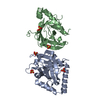
| |||||||||||||||||||||||||||
|---|---|---|---|---|---|---|---|---|---|---|---|---|---|---|---|---|---|---|---|---|---|---|---|---|---|---|---|---|
| 1 | 
| |||||||||||||||||||||||||||
| 2 | 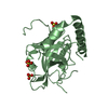
| |||||||||||||||||||||||||||
| Unit cell |
| |||||||||||||||||||||||||||
| Noncrystallographic symmetry (NCS) | NCS domain:
NCS domain segments:
|
-Components
| #1: Protein | Mass: 25775.227 Da / Num. of mol.: 2 / Fragment: UNP residues 43-260 Source method: isolated from a genetically manipulated source Source: (gene. exp.) Streptococcus agalactiae serogroup V (bacteria)Strain: SAG 2603V/R / Gene: SAG0647, srtC1 (SAG0647) / Production host: #2: Chemical | ChemComp-SO4 /   Mass: 96.063 Da / Num. of mol.: 6 / Source method: obtained synthetically / Formula: SO4 Mass: 96.063 Da / Num. of mol.: 6 / Source method: obtained synthetically / Formula: SO4#3: Water | ChemComp-HOH / |  Mass: 18.015 Da / Num. of mol.: 37 / Source method: isolated from a natural source / Formula: H2O Mass: 18.015 Da / Num. of mol.: 37 / Source method: isolated from a natural source / Formula: H2O |
|---|
-Experimental details
-Experiment
| Experiment | Method: X-RAY DIFFRACTION / Number of used crystals: 1 |
|---|
- Sample preparation
| Crystal | Density Matthews: 2.76 Å3/Da / Density % sol: 55.36 % |
|---|---|
| Crystal grow | Temperature: 277 K / Method: vapor diffusion, hanging drop / pH: 6.5 Details: 1.6 M ammonium sulfate, 100 mM calcium chloride, 100 mM sodium cacodylate, pH 6.5, VAPOR DIFFUSION, HANGING DROP, temperature 277K |
-Data collection
| Diffraction | Mean temperature: 100 K |
|---|---|
| Diffraction source | Source: SYNCHROTRON / Site: APS  / Beamline: 22-ID / Wavelength: 1 / Beamline: 22-ID / Wavelength: 1 |
| Detector | Type: MARMOSAIC 300 mm CCD / Detector: CCD / Date: Jul 11, 2007 |
| Radiation | Protocol: SINGLE WAVELENGTH / Monochromatic (M) / Laue (L): M / Scattering type: x-ray |
| Radiation wavelength | Wavelength: 1 Å / Relative weight: 1 |
| Reflection | Resolution: 3→65.22 Å / Num. all: 12057 / Num. obs: 12026 / % possible obs: 99.9 % / Redundancy: 5.7 % / Rmerge(I) obs: 0.088 / Net I/σ(I): 11.3 |
| Reflection shell | Resolution: 3→3.11 Å / Redundancy: 5.6 % / Rmerge(I) obs: 0.248 / Mean I/σ(I) obs: 4.8 / % possible all: 100 |
- Processing
| Software |
| |||||||||||||||||||||||||||||||||||||||||||||||||||||||||||||||||
|---|---|---|---|---|---|---|---|---|---|---|---|---|---|---|---|---|---|---|---|---|---|---|---|---|---|---|---|---|---|---|---|---|---|---|---|---|---|---|---|---|---|---|---|---|---|---|---|---|---|---|---|---|---|---|---|---|---|---|---|---|---|---|---|---|---|---|
| Refinement | Method to determine structure: MOLECULAR REPLACEMENT / Resolution: 3→65.22 Å / Cor.coef. Fo:Fc: 0.929 / Cor.coef. Fo:Fc free: 0.842 / SU B: 16.633 / SU ML: 0.31 / Cross valid method: THROUGHOUT / ESU R Free: 0.475 / Stereochemistry target values: MAXIMUM LIKELIHOOD Details: HYDROGENS HAVE BEEN ADDED IN THE RIDING POSITIONS U VALUES : REFINED INDIVIDUALLY
| |||||||||||||||||||||||||||||||||||||||||||||||||||||||||||||||||
| Solvent computation | Ion probe radii: 0.8 Å / Shrinkage radii: 0.8 Å / VDW probe radii: 1.4 Å / Solvent model: MASK | |||||||||||||||||||||||||||||||||||||||||||||||||||||||||||||||||
| Displacement parameters | Biso mean: 52.346 Å2
| |||||||||||||||||||||||||||||||||||||||||||||||||||||||||||||||||
| Refinement step | Cycle: LAST / Resolution: 3→65.22 Å
| |||||||||||||||||||||||||||||||||||||||||||||||||||||||||||||||||
| Refine LS restraints |
| |||||||||||||||||||||||||||||||||||||||||||||||||||||||||||||||||
| Refine LS restraints NCS | Dom-ID: 1 / Auth asym-ID: A / Ens-ID: 1 / Number: 1377 / Refine-ID: X-RAY DIFFRACTION
| |||||||||||||||||||||||||||||||||||||||||||||||||||||||||||||||||
| LS refinement shell | Resolution: 3→3.078 Å / Total num. of bins used: 20
|
