 Movie
Movie Controller

[English] 日本語
 Yorodumi
- PDB-3r9v: Cocrystal Structure of Proteolytically Truncated Form of IpaD fro... -
+ Open data

- Basic information
| Entry | Database: PDB / ID: 3r9v | ||||||
|---|---|---|---|---|---|---|---|
| Title | Cocrystal Structure of Proteolytically Truncated Form of IpaD from Shigella flexneri Bound to Deoxycholate | ||||||
 Components Components | Invasin ipaD | ||||||
 Keywords Keywords | CELL INVASION / type III secretion system / tip protein / deoxycholate | ||||||
| Function / homology |  Function and homology information Function and homology informationeffector-mediated activation of host programmed cell death by symbiont / extracellular region Similarity search - Function | ||||||
| Biological species |  Shigella flexneri (bacteria) Shigella flexneri (bacteria) | ||||||
| Method |  X-RAY DIFFRACTION / SYNCHROTRON / MOLECULAR REPLACEMENT / Resolution: 1.9 Å X-RAY DIFFRACTION / SYNCHROTRON / MOLECULAR REPLACEMENT / Resolution: 1.9 Å | ||||||
 Authors Authors | Barta, M.L. / Dickenson, N.E. / Picking, W.L. / Picking, W.D. / Geisbrecht, B.V. | ||||||
 Citation Citation | Journal: Proteins / Year: 2012 Title: Identification of the bile salt binding site on IpaD from Shigella flexneri and the influence of ligand binding on IpaD structure. Authors: Barta, M.L. / Guragain, M. / Adam, P. / Dickenson, N.E. / Patil, M. / Geisbrecht, B.V. / Picking, W.L. / Picking, W.D. | ||||||
| History |
|
- Structure visualization
| Structure viewer | Molecule: MolmilJmol/JSmol |
|---|
- Downloads & links
-Download
| PDBx/mmCIF format | 3r9v.cif.gz | 94 KB | Display | PDBx/mmCIF format |
|---|---|---|---|---|
| PDB format | pdb3r9v.ent.gz | 69.1 KB | Display | PDB format |
| PDBx/mmJSON format | 3r9v.json.gz | Tree view | PDBx/mmJSON format | |
| Others |  Other downloads Other downloads |
-Validation report
| Summary document | 3r9v_validation.pdf.gz | 1 MB | Display | wwPDB validaton report |
|---|---|---|---|---|
| Full document | 3r9v_full_validation.pdf.gz | 1 MB | Display | |
| Data in XML | 3r9v_validation.xml.gz | 20 KB | Display | |
| Data in CIF | 3r9v_validation.cif.gz | 27.8 KB | Display | |
| Arichive directory | https://data.pdbj.org/pub/pdb/validation_reports/r9/3r9vftp://data.pdbj.org/pub/pdb/validation_reports/r9/3r9v | HTTPS FTP |
-Related structure data
| Related structure data |  2j0oS S: Starting model for refinement |
|---|---|
| Similar structure data |
-Links
 PDBj
PDBj- Assembly
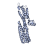
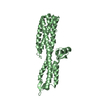
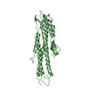
| Deposited unit | 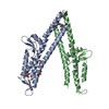
| ||||||||
|---|---|---|---|---|---|---|---|---|---|
| 1 | 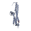
| ||||||||
| 2 | 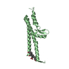
| ||||||||
| 3 |
| ||||||||
| Unit cell |
|
-Components
| #1: Protein | Mass: 31659.391 Da / Num. of mol.: 2 / Fragment: unp residues 39-322 Source method: isolated from a genetically manipulated source Source: (gene. exp.) Shigella flexneri (bacteria) / Gene: ipaD, CP0126 / Production host: #2: Chemical |   Mass: 92.094 Da / Num. of mol.: 3 / Source method: obtained synthetically / Formula: C3H8O3 Mass: 92.094 Da / Num. of mol.: 3 / Source method: obtained synthetically / Formula: C3H8O3#3: Chemical | 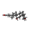  Mass: 392.572 Da / Num. of mol.: 2 / Source method: obtained synthetically / Formula: C24H40O4 / Comment: detergent*YM Mass: 392.572 Da / Num. of mol.: 2 / Source method: obtained synthetically / Formula: C24H40O4 / Comment: detergent*YM#4: Water | ChemComp-HOH / |  Mass: 18.015 Da / Num. of mol.: 226 / Source method: isolated from a natural source / Formula: H2O Mass: 18.015 Da / Num. of mol.: 226 / Source method: isolated from a natural source / Formula: H2O |
|---|
-Experimental details
-Experiment
| Experiment | Method: X-RAY DIFFRACTION / Number of used crystals: 1 |
|---|
- Sample preparation
| Crystal | Density Matthews: 2.02 Å3/Da / Density % sol: 39.15 % |
|---|---|
| Crystal grow | Temperature: 293 K / Method: vapor diffusion, hanging drop / pH: 7.5 Details: 0.02M magnesium chloride hexahydrate, 0.1M HEPES, 22% (w/v) Poly(acrylic acid sodium salt) 5100, pH 7.5, VAPOR DIFFUSION, HANGING DROP, temperature 293K |
-Data collection
| Diffraction | Mean temperature: 100 K |
|---|---|
| Diffraction source | Source: SYNCHROTRON / Site: APS  / Beamline: 22-BM / Wavelength: 1 Å / Beamline: 22-BM / Wavelength: 1 Å |
| Detector | Type: MARMOSAIC 225 mm CCD / Detector: CCD / Date: Feb 6, 2010 |
| Radiation | Monochromator: double crystal - liquid nitrogen cooled / Protocol: SINGLE WAVELENGTH / Monochromatic (M) / Laue (L): M / Scattering type: x-ray |
| Radiation wavelength | Wavelength: 1 Å / Relative weight: 1 |
| Reflection | Resolution: 1.9→45 Å / Num. all: 40352 / Num. obs: 39424 / % possible obs: 97.7 % / Observed criterion σ(F): 0 / Observed criterion σ(I): 2 / Redundancy: 5.7 % / Rsym value: 0.072 / Net I/σ(I): 21.1 |
| Reflection shell | Resolution: 1.9→1.97 Å / Redundancy: 5.7 % / Mean I/σ(I) obs: 2.47 / Rsym value: 0.451 / % possible all: 82.9 |
- Processing
| Software |
| |||||||||||||||||||||||||||||||||||||||||||||||||||||||||||||||||||||||||||||||||||||||||||||||||||||||||
|---|---|---|---|---|---|---|---|---|---|---|---|---|---|---|---|---|---|---|---|---|---|---|---|---|---|---|---|---|---|---|---|---|---|---|---|---|---|---|---|---|---|---|---|---|---|---|---|---|---|---|---|---|---|---|---|---|---|---|---|---|---|---|---|---|---|---|---|---|---|---|---|---|---|---|---|---|---|---|---|---|---|---|---|---|---|---|---|---|---|---|---|---|---|---|---|---|---|---|---|---|---|---|---|---|---|---|
| Refinement | Method to determine structure: MOLECULAR REPLACEMENT Starting model: PDB ENTRY 2J0O Resolution: 1.9→25.283 Å / SU ML: 0.23 / σ(F): 1.35 / Phase error: 26.32 / Stereochemistry target values: ML
| |||||||||||||||||||||||||||||||||||||||||||||||||||||||||||||||||||||||||||||||||||||||||||||||||||||||||
| Solvent computation | Shrinkage radii: 1.06 Å / VDW probe radii: 1.3 Å / Solvent model: FLAT BULK SOLVENT MODEL / Bsol: 41.141 Å2 / ksol: 0.336 e/Å3 | |||||||||||||||||||||||||||||||||||||||||||||||||||||||||||||||||||||||||||||||||||||||||||||||||||||||||
| Displacement parameters |
| |||||||||||||||||||||||||||||||||||||||||||||||||||||||||||||||||||||||||||||||||||||||||||||||||||||||||
| Refinement step | Cycle: LAST / Resolution: 1.9→25.283 Å
| |||||||||||||||||||||||||||||||||||||||||||||||||||||||||||||||||||||||||||||||||||||||||||||||||||||||||
| Refine LS restraints |
| |||||||||||||||||||||||||||||||||||||||||||||||||||||||||||||||||||||||||||||||||||||||||||||||||||||||||
| LS refinement shell |
|
