 Movie
Movie Controller

[English] 日本語
 Yorodumi
- PDB-3r6k: Crystal Structure of the Capsid P Domain from Norwalk Virus Strai... -
+ Open data

- Basic information
| Entry | Database: PDB / ID: 3r6k | |||||||||
|---|---|---|---|---|---|---|---|---|---|---|
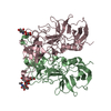
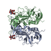
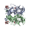
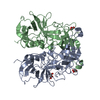
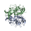
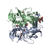
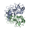
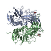
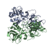
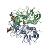
| Title | Crystal Structure of the Capsid P Domain from Norwalk Virus Strain Hiroshima/1999 in complex with HBGA type B (triglycan) | |||||||||
 Components Components | VP1 protein | |||||||||
 Keywords Keywords | VIRAL PROTEIN / Norovirus / P-Domain / Capsid / Receptor / Histo Blood Group Antigen (HBGA) | |||||||||
| Function / homology |  Function and homology information Function and homology information | |||||||||
| Biological species |  Norwalk-like virus Norwalk-like virus | |||||||||
| Method |  X-RAY DIFFRACTION / SYNCHROTRON / MOLECULAR REPLACEMENT / Resolution: 1.6 Å X-RAY DIFFRACTION / SYNCHROTRON / MOLECULAR REPLACEMENT / Resolution: 1.6 Å | |||||||||
 Authors Authors | Hansman, G.S. / Biertumpfel, C. / McLellan, J.S. / Georgiev, I. / Chen, L. / Zhou, T. / Katayama, K. / Kwong, P.D. | |||||||||
 Citation Citation | Journal: J.Virol. / Year: 2011 Title: Crystal structures of GII.10 and GII.12 norovirus protruding domains in complex with histo-blood group antigens reveal details for a potential site of vulnerability. Authors: Hansman, G.S. / Biertumpfel, C. / Georgiev, I. / McLellan, J.S. / Chen, L. / Zhou, T. / Katayama, K. / Kwong, P.D. | |||||||||
| History |
|
- Structure visualization
| Structure viewer | Molecule: MolmilJmol/JSmol |
|---|
- Downloads & links
-Download
| PDBx/mmCIF format | 3r6k.cif.gz | 139.2 KB | Display | PDBx/mmCIF format |
|---|---|---|---|---|
| PDB format | pdb3r6k.ent.gz | 107 KB | Display | PDB format |
| PDBx/mmJSON format | 3r6k.json.gz | Tree view | PDBx/mmJSON format | |
| Others |  Other downloads Other downloads |
-Validation report
| Arichive directory | https://data.pdbj.org/pub/pdb/validation_reports/r6/3r6kftp://data.pdbj.org/pub/pdb/validation_reports/r6/3r6k | HTTPS FTP |
|---|
-Related structure data
| Related structure data | 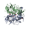 3onuSC  3onyC  3pa1C  3pa2C  3q38C  3q39C  3q3aC  3q6qC  3q6rC  3r6jC C: citing same article ( S: Starting model for refinement |
|---|---|
| Similar structure data |
-Links
 PDBj
PDBj
- Assembly
| Deposited unit | 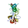
| ||||||||||||
|---|---|---|---|---|---|---|---|---|---|---|---|---|---|
| 1 | 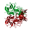
| ||||||||||||
| Unit cell |
| ||||||||||||
| Components on special symmetry positions |
|
-Components
| #1: Protein | Mass: 33414.504 Da / Num. of mol.: 1 / Fragment: unp residues 225-525 Source method: isolated from a genetically manipulated source Source: (gene. exp.) Norwalk-like virus / Strain: Hiroshima/1999/JP / Gene: Capsid protein / Plasmid: MBP-HTSHP / Production host:  | ||
|---|---|---|---|
| #2: Polysaccharide | alpha-L-fucopyranose-(1-2)-[alpha-D-galactopyranose-(1-3)]beta-D-galactopyranose Source method: isolated from a genetically manipulated source | ||
| #3: Chemical |   Mass: 62.068 Da / Num. of mol.: 2 / Source method: obtained synthetically / Formula: C2H6O2 Mass: 62.068 Da / Num. of mol.: 2 / Source method: obtained synthetically / Formula: C2H6O2#4: Water | ChemComp-HOH / |  Mass: 18.015 Da / Num. of mol.: 228 / Source method: isolated from a natural source / Formula: H2O Mass: 18.015 Da / Num. of mol.: 228 / Source method: isolated from a natural source / Formula: H2O |
-Experimental details
-Experiment
| Experiment | Method: X-RAY DIFFRACTION / Number of used crystals: 1 |
|---|
- Sample preparation
| Crystal | Density Matthews: 2.26 Å3/Da / Density % sol: 45.62 % |
|---|---|
| Crystal grow | Temperature: 293 K / Method: vapor diffusion, hanging drop / pH: 6.5 Details: 0.1 M Imidazole, 8.25% (w/v) PEG 8000, 1% (v/v) MPD, pH 6.5, VAPOR DIFFUSION, HANGING DROP, temperature 293K |
-Data collection
| Diffraction | Mean temperature: 95 K |
|---|---|
| Diffraction source | Source: SYNCHROTRON / Site: APS  / Beamline: 22-BM / Wavelength: 1 Å / Beamline: 22-BM / Wavelength: 1 Å |
| Detector | Type: MARMOSAIC 225 mm CCD / Detector: CCD / Date: Sep 12, 2010 |
| Radiation | Monochromator: Si(111) / Protocol: SINGLE WAVELENGTH / Monochromatic (M) / Laue (L): M / Scattering type: x-ray |
| Radiation wavelength | Wavelength: 1 Å / Relative weight: 1 |
| Reflection | Resolution: 1.6→24.446 Å / Num. obs: 37085 / % possible obs: 91.7 % / Observed criterion σ(F): 0 / Observed criterion σ(I): -3 / Redundancy: 5.9 % / Biso Wilson estimate: 22.53 Å2 / Rsym value: 0.086 / Net I/σ(I): 18.26 |
| Reflection shell | Resolution: 1.6→1.66 Å / Redundancy: 3.1 % / Mean I/σ(I) obs: 1.99 / Num. unique all: 2432 / Rsym value: 0.395 / % possible all: 61.2 |
- Processing
| Software |
| ||||||||||||||||||||||||||||||||||||||||||||||||||||||||||||||||||||||||||||||||||||||||||||||||||||
|---|---|---|---|---|---|---|---|---|---|---|---|---|---|---|---|---|---|---|---|---|---|---|---|---|---|---|---|---|---|---|---|---|---|---|---|---|---|---|---|---|---|---|---|---|---|---|---|---|---|---|---|---|---|---|---|---|---|---|---|---|---|---|---|---|---|---|---|---|---|---|---|---|---|---|---|---|---|---|---|---|---|---|---|---|---|---|---|---|---|---|---|---|---|---|---|---|---|---|---|---|---|
| Refinement | Method to determine structure: MOLECULAR REPLACEMENT Starting model: PDB ENTRY 3ONU Resolution: 1.6→24.446 Å / SU ML: 0.19 / Isotropic thermal model: Isotropic, TLS / Cross valid method: THROUGHOUT / σ(F): 0 / Phase error: 27.26 / Stereochemistry target values: ML
| ||||||||||||||||||||||||||||||||||||||||||||||||||||||||||||||||||||||||||||||||||||||||||||||||||||
| Solvent computation | Shrinkage radii: 0.95 Å / VDW probe radii: 1.2 Å / Solvent model: FLAT BULK SOLVENT MODEL / Bsol: 40 Å2 / ksol: 0.31 e/Å3 | ||||||||||||||||||||||||||||||||||||||||||||||||||||||||||||||||||||||||||||||||||||||||||||||||||||
| Displacement parameters |
| ||||||||||||||||||||||||||||||||||||||||||||||||||||||||||||||||||||||||||||||||||||||||||||||||||||
| Refinement step | Cycle: LAST / Resolution: 1.6→24.446 Å
| ||||||||||||||||||||||||||||||||||||||||||||||||||||||||||||||||||||||||||||||||||||||||||||||||||||
| Refine LS restraints |
| ||||||||||||||||||||||||||||||||||||||||||||||||||||||||||||||||||||||||||||||||||||||||||||||||||||
| LS refinement shell |
| ||||||||||||||||||||||||||||||||||||||||||||||||||||||||||||||||||||||||||||||||||||||||||||||||||||
| Refinement TLS params. | Method: refined / Refine-ID: X-RAY DIFFRACTION
| ||||||||||||||||||||||||||||||||||||||||||||||||||||||||||||||||||||||||||||||||||||||||||||||||||||
| Refinement TLS group |
|
