Protocol: SINGLE WAVELENGTH / Scattering type: x-ray
Radiation wavelength
Wavelength: 0.9793 Å / Relative weight: 1
Reflection
Resolution: 1.3→28.9 Å / Num. obs: 28989 / % possible obs: 100 % / Redundancy: 14.2 % / Rmerge(I) obs: 0.049 / Χ2: 0.996 / Net I/σ(I): 12.3
Reflection shell
Resolution (Å)
Redundancy (%)
Rmerge(I) obs
Num. unique all
Χ2
Diffraction-ID
% possible all
1.3-1.32
7.3
0.535
1411
0.915
1
99.9
1.32-1.35
8.6
0.494
1420
0.899
1
100
1.35-1.37
10.2
0.43
1411
0.922
1
100
1.37-1.4
12.6
0.365
1409
0.933
1
100
1.4-1.43
15.4
0.339
1434
0.952
1
100
1.43-1.46
15.4
0.287
1422
0.968
1
100
1.46-1.5
15.5
0.235
1427
1.009
1
100
1.5-1.54
15.5
0.195
1435
1.015
1
100
1.54-1.59
15.5
0.157
1423
1.038
1
100
1.59-1.64
15.6
0.142
1437
1.043
1
100
1.64-1.7
15.6
0.118
1424
1.007
1
100
1.7-1.76
15.6
0.092
1446
1
1
100
1.76-1.84
15.6
0.072
1442
0.966
1
100
1.84-1.94
15.5
0.054
1455
0.922
1
100
1.94-2.06
15.5
0.043
1453
0.889
1
100
2.06-2.22
15.5
0.041
1463
0.935
1
100
2.22-2.45
15.4
0.048
1459
1.341
1
100
2.45-2.8
15.2
0.048
1487
1.584
1
100
2.8-3.53
14.6
0.029
1507
0.859
1
100
3.53-50
14.3
0.023
1624
0.59
1
99.6
-
Processing
Software
Name
Version
Classification
NB
SCALEPACK
datascaling
REFMAC
refinement
PDB_EXTRACT
3.1
dataextraction
CBASS
datacollection
HKL-3000
datareduction
PHASER
phasing
Refinement
Method to determine structure: 1NEU / Resolution: 1.3→28.9 Å / Cor.coef. Fo:Fc: 0.974 / Cor.coef. Fo:Fc free: 0.972 / WRfactor Rfree: 0.172 / WRfactor Rwork: 0.1519 / Occupancy max: 1 / Occupancy min: 0.25 / FOM work R set: 0.9184 / SU B: 1.233 / SU ML: 0.025 / SU R Cruickshank DPI: 0.0576 / SU Rfree: 0.0499 / Cross valid method: THROUGHOUT / σ(F): 0 / ESU R: 0.058 / ESU R Free: 0.05 / Phase error: 26.25 / Stereochemistry target values: MAXIMUM LIKELIHOOD Details: HYDROGENS HAVE BEEN ADDED IN THE RIDING POSITIONS U VALUES : REFINED INDIVIDUALLY : SYMMETRY VIOLATING CLASH FOUND BETWEEN ONE OF THE ROTAMERS (~ 50% OCCUPANCY) OF TRP-155.
Rfactor
Num. reflection
% reflection
Selection details
Rfree
0.1733
1423
5.1 %
RANDOM
Rwork
0.1523
-
-
-
obs
0.1534
28078
97.05 %
-
Solvent computation
Ion probe radii: 0.8 Å / Shrinkage radii: 0.8 Å / VDW probe radii: 1.4 Å / Solvent model: MASK
In the structure databanks used in Yorodumi, some data are registered as the other names, "COVID-19 virus" and "2019-nCoV". Here are the details of the virus and the list of structure data.
Jan 31, 2019. EMDB accession codes are about to change! (news from PDBe EMDB page)
EMDB accession codes are about to change! (news from PDBe EMDB page)
The allocation of 4 digits for EMDB accession codes will soon come to an end. Whilst these codes will remain in use, new EMDB accession codes will include an additional digit and will expand incrementally as the available range of codes is exhausted. The current 4-digit format prefixed with “EMD-” (i.e. EMD-XXXX) will advance to a 5-digit format (i.e. EMD-XXXXX), and so on. It is currently estimated that the 4-digit codes will be depleted around Spring 2019, at which point the 5-digit format will come into force.
The EM Navigator/Yorodumi systems omit the EMD- prefix.
Related info.:Q: What is EMD? / ID/Accession-code notation in Yorodumi/EM Navigator
Yorodumi is a browser for structure data from EMDB, PDB, SASBDB, etc.
This page is also the successor to EM Navigator detail page, and also detail information page/front-end page for Omokage search.
The word "yorodu" (or yorozu) is an old Japanese word meaning "ten thousand". "mi" (miru) is to see.
Related info.:EMDB / PDB / SASBDB / Comparison of 3 databanks / Yorodumi Search / Aug 31, 2016. New EM Navigator & Yorodumi / Yorodumi Papers / Jmol/JSmol / Function and homology information / Changes in new EM Navigator and Yorodumi
 Movie
Movie Controller

 Yorodumi
 Open data
 Basic information
 Components
 Keywords
 Function and homology information
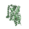
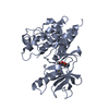
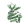
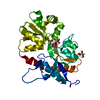
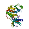
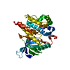
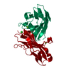
 Homo sapiens (human)
 X-RAY DIFFRACTION /
  Authors
 Citation
 Structure visualization
 Downloads & links
 Other downloads
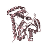


 PDBj
PDBj


 Assembly


 Mass: 24.305 Da / Num. of mol.: 1 / Source method: obtained synthetically / Formula: Mg
 Mass: 35.453 Da / Num. of mol.: 1 / Source method: obtained synthetically / Formula: Cl
 Mass: 18.015 Da / Num. of mol.: 134 / Source method: isolated from a natural source / Formula: H2O
 Sample preparation
 / Beamline: X29A / Wavelength: 0.9793 Å
 Processing