 Movie
Movie Controller


+ Open data

- Basic information
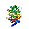
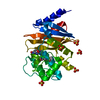
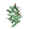
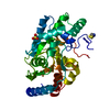
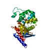
| Entry | Database: PDB / ID: 3qyc | ||||||
|---|---|---|---|---|---|---|---|
| Title | Structure of a dimeric anti-HER2 single domain antibody | ||||||
 Components Components | VH domain of IgG molecule | ||||||
 Keywords Keywords | IMMUNE SYSTEM / immunoglobulin V domain fold / antibody | ||||||
| Function / homology | Immunoglobulins / Immunoglobulin-like / Sandwich / Mainly Beta Function and homology information Function and homology information | ||||||
| Biological species |  Homo sapiens (human) Homo sapiens (human) | ||||||
| Method |  X-RAY DIFFRACTION / SYNCHROTRON / MOLECULAR REPLACEMENT / Resolution: 1.6 Å X-RAY DIFFRACTION / SYNCHROTRON / MOLECULAR REPLACEMENT / Resolution: 1.6 Å | ||||||
 Authors Authors | Baral, T.N. / Chao, S. / Li, S. / Tanha, J. / Arbabai, M. / Wang, S. / Zhang, J. | ||||||
 Citation Citation | Journal: Plos One / Year: 2012 Title: Crystal Structure of a Human Single Domain Antibody Dimer Formed through V(H)-V(H) Non-Covalent Interactions. Authors: Baral, T.N. / Chao, S.Y. / Li, S. / Tanha, J. / Arbabi-Ghahroudi, M. / Zhang, J. / Wang, S. | ||||||
| History |
|
- Structure visualization
| Structure viewer | Molecule: MolmilJmol/JSmol |
|---|
- Downloads & links
-Download
| PDBx/mmCIF format | 3qyc.cif.gz | 59 KB | Display | PDBx/mmCIF format |
|---|---|---|---|---|
| PDB format | pdb3qyc.ent.gz | 46.2 KB | Display | PDB format |
| PDBx/mmJSON format | 3qyc.json.gz | Tree view | PDBx/mmJSON format | |
| Others |  Other downloads Other downloads |
-Validation report
| Summary document | 3qyc_validation.pdf.gz | 423.4 KB | Display | wwPDB validaton report |
|---|---|---|---|---|
| Full document | 3qyc_full_validation.pdf.gz | 425.3 KB | Display | |
| Data in XML | 3qyc_validation.xml.gz | 14 KB | Display | |
| Data in CIF | 3qyc_validation.cif.gz | 20.2 KB | Display | |
| Arichive directory | https://data.pdbj.org/pub/pdb/validation_reports/qy/3qycftp://data.pdbj.org/pub/pdb/validation_reports/qy/3qyc | HTTPS FTP |
-Related structure data
| Similar structure data |
|---|


-Links
 PDBj
PDBj

- Assembly
| Deposited unit | 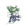
| ||||||||
|---|---|---|---|---|---|---|---|---|---|
| 1 |
| ||||||||
| Unit cell |
|
-Components
| #1: Antibody | Mass: 15740.417 Da / Num. of mol.: 2 Source method: isolated from a genetically manipulated source Source: (gene. exp.) Homo sapiens (human) / Production host:  #2: Water | ChemComp-HOH / |  Mass: 18.015 Da / Num. of mol.: 266 / Source method: isolated from a natural source / Formula: H2O Mass: 18.015 Da / Num. of mol.: 266 / Source method: isolated from a natural source / Formula: H2O |
|---|
-Experimental details
-Experiment
| Experiment | Method: X-RAY DIFFRACTION / Number of used crystals: 1 |
|---|
- Sample preparation
| Crystal | Density Matthews: 1.98 Å3/Da / Density % sol: 37.88 % |
|---|---|
| Crystal grow | Temperature: 298 K / Method: vapor diffusion, sitting drop / pH: 9 Details: 20% PEG 6000, 0.1M Bicine, pH 9.0, VAPOR DIFFUSION, SITTING DROP, temperature 298K |
-Data collection
| Diffraction | Mean temperature: 100 K |
|---|---|
| Diffraction source | Source: SYNCHROTRON / Site: NSRRC  / Beamline: BL13B1 / Wavelength: 1 Å / Beamline: BL13B1 / Wavelength: 1 Å |
| Detector | Type: ADSC QUANTUM 315 / Detector: CCD / Date: Nov 6, 2009 |
| Radiation | Monochromator: GRAPHITE / Protocol: SINGLE WAVELENGTH / Monochromatic (M) / Laue (L): M / Scattering type: x-ray |
| Radiation wavelength | Wavelength: 1 Å / Relative weight: 1 |
| Reflection | Resolution: 1.6→30 Å / Num. all: 33740 / Num. obs: 33598 / % possible obs: 99.4 % / Observed criterion σ(F): 2 / Observed criterion σ(I): 2 / Redundancy: 6.4 % / Rmerge(I) obs: 0.052 |
| Reflection shell | Highest resolution: 1.6 Å / Redundancy: 6.4 % / Rmerge(I) obs: 0.052 / Num. unique all: 33598 / % possible all: 99.4 |
- Processing
| Software |
| ||||||||||||||||||||
|---|---|---|---|---|---|---|---|---|---|---|---|---|---|---|---|---|---|---|---|---|---|
| Refinement | Method to determine structure: MOLECULAR REPLACEMENT / Resolution: 1.6→20 Å / σ(F): 2 / Stereochemistry target values: Engh & Huber
| ||||||||||||||||||||
| Refine analyze |
| ||||||||||||||||||||
| Refinement step | Cycle: LAST / Resolution: 1.6→20 Å
| ||||||||||||||||||||
| Refine LS restraints |
|
