 Movie
Movie Controller

[English] 日本語
 Yorodumi
- PDB-3qwt: The crystal structure of a possible member of GH105 family from S... -
+ Open data

- Basic information
| Entry | Database: PDB / ID: 3qwt | ||||||
|---|---|---|---|---|---|---|---|
| Title | The crystal structure of a possible member of GH105 family from Salmonella enterica subsp. enterica serovar Paratyphi A str. ATCC 9150 | ||||||
 Components Components | Putative GH105 family protein | ||||||
 Keywords Keywords | UNKNOWN FUNCTION / structural genomics / PSI-Biology / protein structure initiative / midwest center for structural genomic s / MCSG / Hydrolase / GH105 / Midwest Center for Structural Genomics / alpha/alpha toroid | ||||||
| Function / homology |  Function and homology information Function and homology informationcatalytic activity / carbohydrate metabolic process / hydrolase activity Similarity search - Function | ||||||
| Biological species |  Salmonella enterica subsp. enterica serovar Paratyphi A (bacteria) Salmonella enterica subsp. enterica serovar Paratyphi A (bacteria) | ||||||
| Method |  X-RAY DIFFRACTION / SYNCHROTRON / MAD / Resolution: 2.183 Å X-RAY DIFFRACTION / SYNCHROTRON / MAD / Resolution: 2.183 Å | ||||||
 Authors Authors | Tan, K. / Hatzos-Skintges, C. / Bearden, J. / Joachimiak, A. / Midwest Center for Structural Genomics (MCSG) | ||||||
 Citation Citation | Journal: To be Published Title: The crystal structure of a possible member of GH105 family from Salmonella enterica subsp. enterica serovar Paratyphi A str. ATCC 9150 Authors: Tan, K. / Hatzos-Skintges, C. / Bearden, J. / Joachimiak, A. | ||||||
| History |
|
- Structure visualization
| Structure viewer | Molecule: MolmilJmol/JSmol |
|---|
- Downloads & links
-Download
| PDBx/mmCIF format | 3qwt.cif.gz | 624.9 KB | Display | PDBx/mmCIF format |
|---|---|---|---|---|
| PDB format | pdb3qwt.ent.gz | 522.9 KB | Display | PDB format |
| PDBx/mmJSON format | 3qwt.json.gz | Tree view | PDBx/mmJSON format | |
| Others |  Other downloads Other downloads |
-Validation report
| Arichive directory | https://data.pdbj.org/pub/pdb/validation_reports/qw/3qwtftp://data.pdbj.org/pub/pdb/validation_reports/qw/3qwt | HTTPS FTP |
|---|
-Related structure data
| Similar structure data | |
|---|---|
| Other databases |

-Links
 PDBj
PDBj
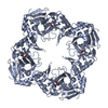
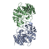
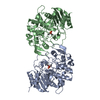
- Assembly
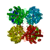
| Deposited unit | 
| ||||||||
|---|---|---|---|---|---|---|---|---|---|
| 1 | 
| ||||||||
| 2 | 
| ||||||||
| Unit cell |
| ||||||||
| Components on special symmetry positions |
| ||||||||
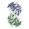
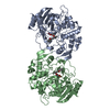
| Details | Experimentally Unknown. It is possilbe the molecule forms dimer. The chains A and B form a dimer. The chain C and its symmetry-related moleucle, by an operator -x,y,-z+1/2, form a dimer. The chain D and its symmtry-related molecule, by an operator, -x,y,-z+1/2, form a dimer. |
-Components
| #1: Protein | Mass: 44912.516 Da / Num. of mol.: 4 Source method: isolated from a genetically manipulated source Source: (gene. exp.) Salmonella enterica subsp. enterica serovar Paratyphi A (bacteria)Gene: SPA0958 / Plasmid: pMCSG7 / Production host: #2: Chemical | ChemComp-GOL /   Mass: 92.094 Da / Num. of mol.: 11 / Source method: obtained synthetically / Formula: C3H8O3 Mass: 92.094 Da / Num. of mol.: 11 / Source method: obtained synthetically / Formula: C3H8O3#3: Water | ChemComp-HOH / |  Mass: 18.015 Da / Num. of mol.: 356 / Source method: isolated from a natural source / Formula: H2O Mass: 18.015 Da / Num. of mol.: 356 / Source method: isolated from a natural source / Formula: H2OHas protein modification | Y | |
|---|
-Experimental details
-Experiment
| Experiment | Method: X-RAY DIFFRACTION / Number of used crystals: 1 |
|---|
- Sample preparation
| Crystal | Density Matthews: 2.4 Å3/Da / Density % sol: 48.83 % |
|---|---|
| Crystal grow | Temperature: 289 K / Method: vapor diffusion, sitting drop / pH: 8.5 Details: 0.2M NaCl, 0.1M Tris.HCl, 25% (w/v) PEG3350, pH 8.5, VAPOR DIFFUSION, SITTING DROP, temperature 289K |
-Data collection
| Diffraction | Mean temperature: 100 K | |||||||||
|---|---|---|---|---|---|---|---|---|---|---|
| Diffraction source | Source: SYNCHROTRON / Site: APS  / Beamline: 19-ID / Wavelength: 0.97942, 0.97951 / Beamline: 19-ID / Wavelength: 0.97942, 0.97951 | |||||||||
| Detector | Type: ADSC QUANTUM 315r / Detector: CCD / Date: Oct 31, 2010 / Details: mirror | |||||||||
| Radiation | Monochromator: Si 111 crystal / Protocol: MAD / Monochromatic (M) / Laue (L): M / Scattering type: x-ray | |||||||||
| Radiation wavelength |
| |||||||||
| Reflection | Resolution: 2.18→40 Å / Num. all: 87941 / Num. obs: 87941 / % possible obs: 98.2 % / Observed criterion σ(F): 0 / Observed criterion σ(I): 0 / Redundancy: 4.5 % / Rmerge(I) obs: 0.107 / Net I/σ(I): 20.7 | |||||||||
| Reflection shell | Resolution: 2.18→2.22 Å / Redundancy: 4.4 % / Rmerge(I) obs: 0.696 / Mean I/σ(I) obs: 2.02 / Num. unique all: 4188 / % possible all: 94.9 |
- Processing
| Software |
| |||||||||||||||||||||||||||||||||||||||||||||||||||||||||||||||||||||||||||||||||||||||||||||||||||||||||||||||||||||||||||||
|---|---|---|---|---|---|---|---|---|---|---|---|---|---|---|---|---|---|---|---|---|---|---|---|---|---|---|---|---|---|---|---|---|---|---|---|---|---|---|---|---|---|---|---|---|---|---|---|---|---|---|---|---|---|---|---|---|---|---|---|---|---|---|---|---|---|---|---|---|---|---|---|---|---|---|---|---|---|---|---|---|---|---|---|---|---|---|---|---|---|---|---|---|---|---|---|---|---|---|---|---|---|---|---|---|---|---|---|---|---|---|---|---|---|---|---|---|---|---|---|---|---|---|---|---|---|---|
| Refinement | Method to determine structure: MAD / Resolution: 2.183→39.84 Å / SU ML: 0.35 / σ(F): 0 / Stereochemistry target values: ML
| |||||||||||||||||||||||||||||||||||||||||||||||||||||||||||||||||||||||||||||||||||||||||||||||||||||||||||||||||||||||||||||
| Solvent computation | Shrinkage radii: 0.83 Å / VDW probe radii: 1.1 Å / Solvent model: FLAT BULK SOLVENT MODEL / Bsol: 40.937 Å2 / ksol: 0.329 e/Å3 | |||||||||||||||||||||||||||||||||||||||||||||||||||||||||||||||||||||||||||||||||||||||||||||||||||||||||||||||||||||||||||||
| Displacement parameters |
| |||||||||||||||||||||||||||||||||||||||||||||||||||||||||||||||||||||||||||||||||||||||||||||||||||||||||||||||||||||||||||||
| Refinement step | Cycle: LAST / Resolution: 2.183→39.84 Å
| |||||||||||||||||||||||||||||||||||||||||||||||||||||||||||||||||||||||||||||||||||||||||||||||||||||||||||||||||||||||||||||
| Refine LS restraints |
| |||||||||||||||||||||||||||||||||||||||||||||||||||||||||||||||||||||||||||||||||||||||||||||||||||||||||||||||||||||||||||||
| LS refinement shell |
| |||||||||||||||||||||||||||||||||||||||||||||||||||||||||||||||||||||||||||||||||||||||||||||||||||||||||||||||||||||||||||||
| Refinement TLS params. | Method: refined / Refine-ID: X-RAY DIFFRACTION
| |||||||||||||||||||||||||||||||||||||||||||||||||||||||||||||||||||||||||||||||||||||||||||||||||||||||||||||||||||||||||||||
| Refinement TLS group |
|
