Resolution: 2.39→2.52 Å / Redundancy: 3.5 % / Rmerge(I) obs: 0.667 / Mean I/σ(I) obs: 2 / Num. unique all: 1343 / % possible all: 99.9
-
Processing
Software
Name
Version
Classification
GDA
datacollection
SHARP
phasing
REFMAC
5.6.0066
refinement
MOSFLM
datareduction
SCALA
datascaling
Refinement
Method to determine structure: SIRAS / Resolution: 2.39→48.07 Å / Cor.coef. Fo:Fc: 0.944 / Cor.coef. Fo:Fc free: 0.929 / SU B: 16.577 / SU ML: 0.176 / Cross valid method: THROUGHOUT / ESU R Free: 0.225 / Stereochemistry target values: MAXIMUM LIKELIHOOD / Details: HYDROGENS HAVE BEEN USED IF PRESENT IN THE INPUT
Rfactor
Num. reflection
% reflection
Selection details
Rfree
0.27242
487
5.1 %
RANDOM
Rwork
0.24228
-
-
-
all
0.24369
9587
-
-
obs
0.24369
9587
99.67 %
-
Solvent computation
Ion probe radii: 0.8 Å / Shrinkage radii: 0.8 Å / VDW probe radii: 1.2 Å / Solvent model: MASK
Displacement parameters
Biso mean: 59.584 Å2
Baniso -1
Baniso -2
Baniso -3
1-
0.5 Å2
0.25 Å2
0 Å2
2-
-
0.5 Å2
0 Å2
3-
-
-
-0.76 Å2
Refinement step
Cycle: LAST / Resolution: 2.39→48.07 Å
Protein
Nucleic acid
Ligand
Solvent
Total
Num. atoms
1035
0
0
16
1051
Refine LS restraints
Refine-ID
Type
Dev ideal
Dev ideal target
Number
X-RAY DIFFRACTION
r_bond_refined_d
0.011
0.022
1056
X-RAY DIFFRACTION
r_bond_other_d
0.001
0.02
736
X-RAY DIFFRACTION
r_angle_refined_deg
1.288
1.966
1427
X-RAY DIFFRACTION
r_angle_other_deg
0.772
3
1783
X-RAY DIFFRACTION
r_dihedral_angle_1_deg
5.854
5
125
X-RAY DIFFRACTION
r_dihedral_angle_2_deg
34.685
23.929
56
X-RAY DIFFRACTION
r_dihedral_angle_3_deg
17.601
15
184
X-RAY DIFFRACTION
r_dihedral_angle_4_deg
18.157
15
9
X-RAY DIFFRACTION
r_chiral_restr
0.074
0.2
157
X-RAY DIFFRACTION
r_gen_planes_refined
0.005
0.021
1166
X-RAY DIFFRACTION
r_gen_planes_other
0.001
0.02
223
X-RAY DIFFRACTION
r_nbd_refined
X-RAY DIFFRACTION
r_nbd_other
X-RAY DIFFRACTION
r_nbtor_refined
X-RAY DIFFRACTION
r_nbtor_other
X-RAY DIFFRACTION
r_xyhbond_nbd_refined
X-RAY DIFFRACTION
r_xyhbond_nbd_other
X-RAY DIFFRACTION
r_metal_ion_refined
X-RAY DIFFRACTION
r_metal_ion_other
X-RAY DIFFRACTION
r_symmetry_vdw_refined
X-RAY DIFFRACTION
r_symmetry_vdw_other
X-RAY DIFFRACTION
r_symmetry_hbond_refined
X-RAY DIFFRACTION
r_symmetry_hbond_other
X-RAY DIFFRACTION
r_symmetry_metal_ion_refined
X-RAY DIFFRACTION
r_symmetry_metal_ion_other
X-RAY DIFFRACTION
r_mcbond_it
X-RAY DIFFRACTION
r_mcbond_other
X-RAY DIFFRACTION
r_mcangle_it
X-RAY DIFFRACTION
r_scbond_it
X-RAY DIFFRACTION
r_scangle_it
X-RAY DIFFRACTION
r_rigid_bond_restr
X-RAY DIFFRACTION
r_sphericity_free
X-RAY DIFFRACTION
r_sphericity_bonded
LS refinement shell
Resolution: 2.39→2.452 Å / Total num. of bins used: 20
Rfactor
Num. reflection
% reflection
Rfree
0.456
32
-
Rwork
0.372
650
-
obs
-
-
99.27 %
Refinement TLS params.
Method: refined / Refine-ID: X-RAY DIFFRACTION
ID
L11 (°2)
L12 (°2)
L13 (°2)
L22 (°2)
L23 (°2)
L33 (°2)
S11 (Å °)
S12 (Å °)
S13 (Å °)
S21 (Å °)
S22 (Å °)
S23 (Å °)
S31 (Å °)
S32 (Å °)
S33 (Å °)
T11 (Å2)
T12 (Å2)
T13 (Å2)
T22 (Å2)
T23 (Å2)
T33 (Å2)
Origin x (Å)
Origin y (Å)
Origin z (Å)
1
0.4467
0.4157
-0.0001
1.0613
-0.1958
0.6111
-0.0503
-0.1914
-0.0758
-0.0155
-0.1348
0.2098
-0.034
0.178
0.1851
0.1397
-0.0469
0.0673
0.1817
0.1091
0.2726
22.4891
28.8338
22.9879
2
0.1507
-0.5048
0.1923
2.2507
-0.0788
0.8675
-0.0149
0.0739
-0.0178
-0.1571
-0.0662
0.1375
-0.1773
0.3677
0.0812
0.1286
-0.0927
0.0305
0.2538
0.0449
0.0788
30.1633
37.1705
19.8883
3
0.0725
0.0647
-0.3563
1.9292
0.4009
2.5914
0.0246
-0.1029
-0.0091
-0.1218
-0.03
-0.0652
0.0112
0.3587
0.0054
0.1042
-0.0546
0.0449
0.22
0.0582
0.0906
32.721
33.9541
17.0138
4
2.0478
0.2043
3.3259
3.0018
-6.7664
26.1334
-0.3573
-0.1212
-0.1251
-0.2364
-0.4313
-0.0114
1.1509
-0.5132
0.7886
0.492
-0.2596
0.3145
0.4218
-0.3211
0.3023
31.5519
29.2678
-5.4461
Refinement TLS group
ID
Refine-ID
Refine TLS-ID
Auth asym-ID
Auth seq-ID
1
X-RAY DIFFRACTION
1
A
736 - 788
2
X-RAY DIFFRACTION
2
A
789 - 812
3
X-RAY DIFFRACTION
3
A
813 - 848
4
X-RAY DIFFRACTION
4
A
849 - 865
+
About Yorodumi
-
News
-
Feb 9, 2022. New format data for meta-information of EMDB entries
New format data for meta-information of EMDB entries
Version 3 of the EMDB header file is now the official format.
The previous official version 1.9 will be removed from the archive.
In the structure databanks used in Yorodumi, some data are registered as the other names, "COVID-19 virus" and "2019-nCoV". Here are the details of the virus and the list of structure data.
Jan 31, 2019. EMDB accession codes are about to change! (news from PDBe EMDB page)
EMDB accession codes are about to change! (news from PDBe EMDB page)
The allocation of 4 digits for EMDB accession codes will soon come to an end. Whilst these codes will remain in use, new EMDB accession codes will include an additional digit and will expand incrementally as the available range of codes is exhausted. The current 4-digit format prefixed with “EMD-” (i.e. EMD-XXXX) will advance to a 5-digit format (i.e. EMD-XXXXX), and so on. It is currently estimated that the 4-digit codes will be depleted around Spring 2019, at which point the 5-digit format will come into force.
The EM Navigator/Yorodumi systems omit the EMD- prefix.
Related info.:Q: What is EMD? / ID/Accession-code notation in Yorodumi/EM Navigator
Yorodumi is a browser for structure data from EMDB, PDB, SASBDB, etc.
This page is also the successor to EM Navigator detail page, and also detail information page/front-end page for Omokage search.
The word "yorodu" (or yorozu) is an old Japanese word meaning "ten thousand". "mi" (miru) is to see.
Related info.:EMDB / PDB / SASBDB / Comparison of 3 databanks / Yorodumi Search / Aug 31, 2016. New EM Navigator & Yorodumi / Yorodumi Papers / Jmol/JSmol / Function and homology information / Changes in new EM Navigator and Yorodumi
 Movie
Movie Controller

 Yorodumi
 Open data
 Basic information
 Components
 Keywords
 Function and homology information
 Homo sapiens (human)
 X-RAY DIFFRACTION /
  Authors
 Citation
 Structure visualization
 Downloads & links
 Other downloads
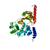

 PDBj
PDBj

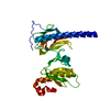
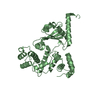
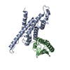
 Assembly

 Mass: 18.015 Da / Num. of mol.: 16 / Source method: isolated from a natural source / Formula: H2O
 Sample preparation
 Processing