 Movie
Movie Controller

[English] 日本語
 Yorodumi
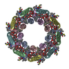
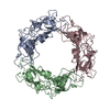
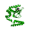
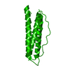
- PDB-1kzu: INTEGRAL MEMBRANE PERIPHERAL LIGHT HARVESTING COMPLEX FROM RHODOP... -
+ Open data

- Basic information
| Entry | Database: PDB / ID: 1kzu | |||||||||
|---|---|---|---|---|---|---|---|---|---|---|
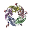
| Title | INTEGRAL MEMBRANE PERIPHERAL LIGHT HARVESTING COMPLEX FROM RHODOPSEUDOMONAS ACIDOPHILA STRAIN 10050 | |||||||||
 Components Components | (LIGHT HARVESTING PROTEIN B-800/850) x 2 | |||||||||
 Keywords Keywords | LIGHT-HARVESTING PROTEIN / ANTENNA COMPLEX / LH2 COMPLEX / BACTERIOCHLOROPHYLL / PURPLE BACTERIA | |||||||||
| Function / homology |  Function and homology information Function and homology informationorganelle inner membrane / plasma membrane light-harvesting complex / bacteriochlorophyll binding / photosynthesis, light reaction / metal ion binding / plasma membrane Similarity search - Function | |||||||||
| Biological species |  Rhodoblastus acidophilus (bacteria) Rhodoblastus acidophilus (bacteria) | |||||||||
| Method |  X-RAY DIFFRACTION / SYNCHROTRON / MIRAS / Resolution: 2.5 Å X-RAY DIFFRACTION / SYNCHROTRON / MIRAS / Resolution: 2.5 Å | |||||||||
 Authors Authors | Cogdell, R.J. / Freer, A.A. / Isaacs, N.W. / Hawthornthwaite-Lawless, A.M. / Mcdermott, G. / Papiz, M.Z. / Prince, S.M. | |||||||||
 Citation Citation | Journal: J.Mol.Biol. / Year: 1997 Title: Apoprotein structure in the LH2 complex from Rhodopseudomonas acidophila strain 10050: modular assembly and protein pigment interactions. Authors: Prince, S.M. / Papiz, M.Z. / Freer, A.A. / McDermott, G. / Hawthornthwaite-Lawless, A.M. / Cogdell, R.J. / Isaacs, N.W. #1: Journal: Photochem.Photobiol. / Year: 1996Title: Structure-Based Calculations on the Optical Spectra of the Lh2 Bacteriochlorophyll-Protein Complex from Rhodopseudomonas Acidophila Authors: Sauer, K. / Cogdell, R.J. / Prince, S.M. / Freer, A. / Isaacs, N.W. / Scheer, H. #2: Journal: Structure / Year: 1996Title: Pigment-Pigment Interactions and Energy Transfer in the Antenna Complex of the Photosynthetic Bacterium Rhodopseudomonas Acidophila Authors: Freer, A. / Prince, S. / Sauer, K. / Papiz, M. / Hawthornthwaite-Lawless, A. / Mcdermott, G. / Cogdell, R. / Isaacs, N.W. #3: Journal: Trends Plant Sci. / Year: 1996Title: A Model for the Photosynthetic Apparatus of Purple Bacteria Authors: Papiz, M.Z. / Prince, S.M. / Hawthornthwaite-Lawless, A.M. / Mcdermott, G. / Freer, A. / Isaacs, N.W. / Cogdell, R.J. #4: Journal: Nature / Year: 1995Title: Crystal Structure of an Integral Membrane Light-Harvesting Complex from Photosynthetic Bacteria Authors: Mcdermott, G. / Prince, S.M. / Freer, A.A. / Hawthornthwaite-Lawless, A.M. / Papiz, M.Z. / Cogdell, R.J. / Isaacs, N.W. #5: Journal: Curr.Opin.Struct.Biol. / Year: 1995Title: Light-Harvesting Mechanisms in Purple Photosynthetic Bacteria Authors: Isaacs, N.W. / Cogdell, R.J. / Freer, A.A. / Prince, S.M. | |||||||||
| History |
|
- Structure visualization
| Structure viewer | Molecule: MolmilJmol/JSmol |
|---|
- Downloads & links
-Download
| PDBx/mmCIF format | 1kzu.cif.gz | 88.2 KB | Display | PDBx/mmCIF format |
|---|---|---|---|---|
| PDB format | pdb1kzu.ent.gz | 69.4 KB | Display | PDB format |
| PDBx/mmJSON format | 1kzu.json.gz | Tree view | PDBx/mmJSON format | |
| Others |  Other downloads Other downloads |
-Validation report
| Arichive directory | https://data.pdbj.org/pub/pdb/validation_reports/kz/1kzuftp://data.pdbj.org/pub/pdb/validation_reports/kz/1kzu | HTTPS FTP |
|---|
-Related structure data
| Similar structure data |
|---|
-Links
 PDBj
PDBj- Assembly
| Deposited unit | 
| |||||||||
|---|---|---|---|---|---|---|---|---|---|---|
| 1 | 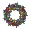
| |||||||||
| Unit cell |
| |||||||||
| Components on special symmetry positions |
| |||||||||
| Noncrystallographic symmetry (NCS) | NCS oper:
| |||||||||
| Details | THE CELL IS A HEXAGONALLY INDEXED RHOMBOHEDRAL CELL. THE DEPOSITORS PROVIDED ONE-THIRD OF THE ASYMMETRIC UNIT. THE REMAINING TWO-THIRDS WERE GENERATED BY THE PDB USING THE MTRIX TRANSFORMATIONS SO THAT THE ENTRY CONTAINS A COMPLETE ASYMMETRIC UNIT. ASSOCIATION OF CHAIN IDENTIFIERS AND HET ATOMS PROTEIN CHAIN HET AND SOLVENT CHAIN A, B C D, E F G, H I |
-Components
| #1: Protein | Mass: 5686.733 Da / Num. of mol.: 3 / Source method: isolated from a natural source / Source: (natural) Rhodoblastus acidophilus (bacteria) / Strain: 10050 / References: UniProt: P26789#2: Protein/peptide | Mass: 4559.203 Da / Num. of mol.: 3 / Source method: isolated from a natural source / Source: (natural) Rhodoblastus acidophilus (bacteria) / Strain: 10050 / References: UniProt: P26790#3: Sugar | ChemComp-RG1 /   Type: D-saccharide / Mass: 715.013 Da / Num. of mol.: 6 Type: D-saccharide / Mass: 715.013 Da / Num. of mol.: 6Source method: isolated from a genetically manipulated source Formula: C46H66O6 #4: Chemical | ChemComp-BCL /   Mass: 911.504 Da / Num. of mol.: 9 / Source method: obtained synthetically / Formula: C55H74MgN4O6 Mass: 911.504 Da / Num. of mol.: 9 / Source method: obtained synthetically / Formula: C55H74MgN4O6#5: Water | ChemComp-HOH / |  Mass: 18.015 Da / Num. of mol.: 51 / Source method: isolated from a natural source / Formula: H2O Mass: 18.015 Da / Num. of mol.: 51 / Source method: isolated from a natural source / Formula: H2OHas protein modification | Y | |
|---|
-Experimental details
-Experiment
| Experiment | Method: X-RAY DIFFRACTION / Number of used crystals: 9 |
|---|
- Sample preparation
| Crystal | Density % sol: 73 % | ||||||||||||||||||||
|---|---|---|---|---|---|---|---|---|---|---|---|---|---|---|---|---|---|---|---|---|---|
| Crystal grow | *PLUS Temperature: 20 ℃ / pH: 9.5 / Method: vapor diffusion | ||||||||||||||||||||
| Components of the solutions | *PLUS
|
-Data collection
| Diffraction | Mean temperature: 300 K |
|---|---|
| Diffraction source | Source: SYNCHROTRON / Site: SRS  / Beamline: PX9.6 / Wavelength: 0.87 / Beamline: PX9.6 / Wavelength: 0.87 |
| Detector | Type: MAR scanner 300 mm plate / Detector: IMAGE PLATE / Date: Dec 1, 1994 |
| Radiation | Monochromatic (M) / Laue (L): M / Scattering type: x-ray |
| Radiation wavelength | Wavelength: 0.87 Å / Relative weight: 1 |
| Reflection | Resolution: 2.5→12 Å / Num. obs: 28574 / % possible obs: 98.9 % / Redundancy: 2.3 % / Biso Wilson estimate: 57.77 Å2 / Rmerge(I) obs: 0.05 / Net I/σ(I): 5.9 |
| Reflection shell | Resolution: 2.5→2.56 Å / Redundancy: 1.8 % / Rmerge(I) obs: 0.239 / Mean I/σ(I) obs: 1.9 / % possible all: 100 |
| Reflection shell | *PLUS % possible obs: 100 % |
- Processing
| Software |
| ||||||||||||||||||||||||||||||||||||||||||||||||||||||||||||||||||||||||||||||||
|---|---|---|---|---|---|---|---|---|---|---|---|---|---|---|---|---|---|---|---|---|---|---|---|---|---|---|---|---|---|---|---|---|---|---|---|---|---|---|---|---|---|---|---|---|---|---|---|---|---|---|---|---|---|---|---|---|---|---|---|---|---|---|---|---|---|---|---|---|---|---|---|---|---|---|---|---|---|---|---|---|---|
| Refinement | Method to determine structure: MIRAS / Resolution: 2.5→12 Å / Rfactor Rfree error: 0.028 / σ(F): 2
| ||||||||||||||||||||||||||||||||||||||||||||||||||||||||||||||||||||||||||||||||
| Displacement parameters | Biso mean: 34.52 Å2
| ||||||||||||||||||||||||||||||||||||||||||||||||||||||||||||||||||||||||||||||||
| Refine analyze |
| ||||||||||||||||||||||||||||||||||||||||||||||||||||||||||||||||||||||||||||||||
| Refinement step | Cycle: LAST / Resolution: 2.5→12 Å
| ||||||||||||||||||||||||||||||||||||||||||||||||||||||||||||||||||||||||||||||||
| Refine LS restraints |
| ||||||||||||||||||||||||||||||||||||||||||||||||||||||||||||||||||||||||||||||||
| Refine LS restraints NCS | NCS model details: CONSTRAINED | ||||||||||||||||||||||||||||||||||||||||||||||||||||||||||||||||||||||||||||||||
| LS refinement shell | Resolution: 2.5→2.61 Å / Rfactor Rfree error: 0.085
| ||||||||||||||||||||||||||||||||||||||||||||||||||||||||||||||||||||||||||||||||
| Xplor file |
| ||||||||||||||||||||||||||||||||||||||||||||||||||||||||||||||||||||||||||||||||
| Software | *PLUS Name: X-PLOR / Classification: refinement | ||||||||||||||||||||||||||||||||||||||||||||||||||||||||||||||||||||||||||||||||
| Refinement | *PLUS Num. reflection obs: 27855 / Rfactor Rfree: 0.253 | ||||||||||||||||||||||||||||||||||||||||||||||||||||||||||||||||||||||||||||||||
| Solvent computation | *PLUS | ||||||||||||||||||||||||||||||||||||||||||||||||||||||||||||||||||||||||||||||||
| Displacement parameters | *PLUS | ||||||||||||||||||||||||||||||||||||||||||||||||||||||||||||||||||||||||||||||||
| Refine LS restraints | *PLUS
| ||||||||||||||||||||||||||||||||||||||||||||||||||||||||||||||||||||||||||||||||
| LS refinement shell | *PLUS Rfactor obs: 0.302 |
