 Movie
Movie Controller

[English] 日本語
 Yorodumi
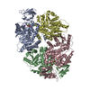
- PDB-3qv8: Crystal structure of Leishmania mexicana pyruvate kinase(LmPYK)in... -
+ Open data

- Basic information
| Entry | Database: PDB / ID: 3qv8 | ||||||
|---|---|---|---|---|---|---|---|
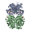
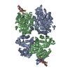
| Title | Crystal structure of Leishmania mexicana pyruvate kinase(LmPYK)in complex with benzothiazole-2,5-disulfonic acid. | ||||||
 Components Components | Pyruvate kinase | ||||||
 Keywords Keywords | TRANSFERASE/TRANSFERASE INHIBITOR / PYRUVATE KINASE / GLYCOLYSIS / TIM Barrel / Sugar kinase / ADP/ATP binding / cytosol / TRANSFERASE-TRANSFERASE INHIBITOR complex | ||||||
| Function / homology |  Function and homology information Function and homology informationpyruvate kinase / pyruvate kinase activity / potassium ion binding / response to stress / kinase activity / magnesium ion binding / ATP binding Similarity search - Function | ||||||
| Biological species |   Leishmania mexicana (eukaryote) Leishmania mexicana (eukaryote) | ||||||
| Method |  X-RAY DIFFRACTION / SYNCHROTRON / MOLECULAR REPLACEMENT / Resolution: 2.45 Å X-RAY DIFFRACTION / SYNCHROTRON / MOLECULAR REPLACEMENT / Resolution: 2.45 Å | ||||||
 Authors Authors | Morgan, H.P. / Auld, D.S. / McNae, I.W. / Nowicki, M.W. / Michels, P.A.M. / Fothergill-Gilmore, L.A. / Walkinshaw, M.D. | ||||||
 Citation Citation | Journal: J.Biol.Chem. / Year: 2011 Title: The trypanocidal drug suramin and other trypan blue mimetics are inhibitors of pyruvate kinases and bind to the adenosine site. Authors: Morgan, H.P. / McNae, I.W. / Nowicki, M.W. / Zhong, W. / Michels, P.A. / Auld, D.S. / Fothergill-Gilmore, L.A. / Walkinshaw, M.D. | ||||||
| History |
|
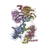
- Structure visualization
| Structure viewer | Molecule: MolmilJmol/JSmol |
|---|
- Downloads & links
-Download
| PDBx/mmCIF format | 3qv8.cif.gz | 182.9 KB | Display | PDBx/mmCIF format |
|---|---|---|---|---|
| PDB format | pdb3qv8.ent.gz | 145.4 KB | Display | PDB format |
| PDBx/mmJSON format | 3qv8.json.gz | Tree view | PDBx/mmJSON format | |
| Others |  Other downloads Other downloads |
-Validation report
| Arichive directory | https://data.pdbj.org/pub/pdb/validation_reports/qv/3qv8ftp://data.pdbj.org/pub/pdb/validation_reports/qv/3qv8 | HTTPS FTP |
|---|
-Related structure data
| Related structure data |  3pp7C 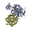 3qv6C  3qv7C  3qv9C  1pklS S: Starting model for refinement C: citing same article ( |
|---|---|
| Similar structure data |


-Links
 PDBj
PDBj
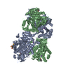
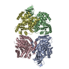
- Assembly
| Deposited unit | 
| ||||||||
|---|---|---|---|---|---|---|---|---|---|
| 1 | 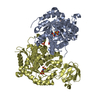
| ||||||||
| Unit cell |
|
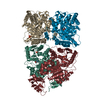
-Components
| #1: Protein | Mass: 54467.934 Da / Num. of mol.: 2 Source method: isolated from a genetically manipulated source Source: (gene. exp.) Leishmania mexicana (eukaryote) / Strain: BEL46 / Gene: PKY / Plasmid: pET3a / Production host:  #2: Chemical |   Mass: 295.313 Da / Num. of mol.: 2 / Source method: obtained synthetically / Formula: C7H5NO6S3 Mass: 295.313 Da / Num. of mol.: 2 / Source method: obtained synthetically / Formula: C7H5NO6S3#3: Chemical | ChemComp-PO4 /   Mass: 94.971 Da / Num. of mol.: 4 / Source method: obtained synthetically / Formula: PO4 Mass: 94.971 Da / Num. of mol.: 4 / Source method: obtained synthetically / Formula: PO4#4: Water | ChemComp-HOH / |  Mass: 18.015 Da / Num. of mol.: 211 / Source method: isolated from a natural source / Formula: H2O Mass: 18.015 Da / Num. of mol.: 211 / Source method: isolated from a natural source / Formula: H2O |
|---|
-Experimental details
-Experiment
| Experiment | Method: X-RAY DIFFRACTION |
|---|
- Sample preparation
| Crystal | Density Matthews: 3.04 Å3/Da / Density % sol: 59.59 % |
|---|---|
| Crystal grow | Temperature: 277 K / Method: vapor diffusion, hanging drop / pH: 7.2 Details: 7-16% PEG 8,000, 20 mM TEA buffer (pH 7.2), 50 mM MgCl2, 100 mM KCl and 10-25% glycerol, VAPOR DIFFUSION, HANGING DROP, temperature 277K |
-Data collection
| Diffraction source | Source: SYNCHROTRON / Site: Diamond  / Beamline: I03 / Wavelength: 1 Å / Beamline: I03 / Wavelength: 1 Å | ||||||||||||||||||
|---|---|---|---|---|---|---|---|---|---|---|---|---|---|---|---|---|---|---|---|
| Detector | Type: ADSC QUANTUM 315 / Detector: CCD | ||||||||||||||||||
| Radiation | Protocol: SINGLE WAVELENGTH / Monochromatic (M) / Laue (L): M / Scattering type: x-ray | ||||||||||||||||||
| Radiation wavelength | Wavelength: 1 Å / Relative weight: 1 | ||||||||||||||||||
| Reflection | Resolution: 2.45→44.41 Å / Num. all: 360184 / Num. obs: 49123 / % possible obs: 100 % / Observed criterion σ(F): 2.45 / Observed criterion σ(I): 2.45 / Redundancy: 7.3 % / Biso Wilson estimate: 52.1 Å2 / Rmerge(I) obs: 0.095 / Rsym value: 0.102 / Net I/σ(I): 16.1 | ||||||||||||||||||
| Reflection shell | Diffraction-ID: 1 / % possible all: 100
|
- Processing
| Software |
| ||||||||||||||||||||||||||||||||||||||||||||||||||||||||||||||||||||||||||||||||||||||||||||||||||||||||||||||||||||||||||||||||||||||||||||||||||||||||||||||||||||||||||
|---|---|---|---|---|---|---|---|---|---|---|---|---|---|---|---|---|---|---|---|---|---|---|---|---|---|---|---|---|---|---|---|---|---|---|---|---|---|---|---|---|---|---|---|---|---|---|---|---|---|---|---|---|---|---|---|---|---|---|---|---|---|---|---|---|---|---|---|---|---|---|---|---|---|---|---|---|---|---|---|---|---|---|---|---|---|---|---|---|---|---|---|---|---|---|---|---|---|---|---|---|---|---|---|---|---|---|---|---|---|---|---|---|---|---|---|---|---|---|---|---|---|---|---|---|---|---|---|---|---|---|---|---|---|---|---|---|---|---|---|---|---|---|---|---|---|---|---|---|---|---|---|---|---|---|---|---|---|---|---|---|---|---|---|---|---|---|---|---|---|---|---|
| Refinement | Method to determine structure: MOLECULAR REPLACEMENT Starting model: 1PKL Resolution: 2.45→44.41 Å / Cor.coef. Fo:Fc: 0.931 / Cor.coef. Fo:Fc free: 0.915 / SU B: 8.583 / SU ML: 0.194 / Cross valid method: THROUGHOUT / ESU R Free: 0.258 / Stereochemistry target values: MAXIMUM LIKELIHOOD / Details: HYDROGENS HAVE BEEN ADDED IN THE RIDING POSITIONS
| ||||||||||||||||||||||||||||||||||||||||||||||||||||||||||||||||||||||||||||||||||||||||||||||||||||||||||||||||||||||||||||||||||||||||||||||||||||||||||||||||||||||||||
| Solvent computation | Ion probe radii: 0.8 Å / Shrinkage radii: 0.8 Å / VDW probe radii: 1.2 Å / Solvent model: BABINET MODEL WITH MASK | ||||||||||||||||||||||||||||||||||||||||||||||||||||||||||||||||||||||||||||||||||||||||||||||||||||||||||||||||||||||||||||||||||||||||||||||||||||||||||||||||||||||||||
| Displacement parameters | Biso mean: 52.196 Å2
| ||||||||||||||||||||||||||||||||||||||||||||||||||||||||||||||||||||||||||||||||||||||||||||||||||||||||||||||||||||||||||||||||||||||||||||||||||||||||||||||||||||||||||
| Refinement step | Cycle: LAST / Resolution: 2.45→44.41 Å
| ||||||||||||||||||||||||||||||||||||||||||||||||||||||||||||||||||||||||||||||||||||||||||||||||||||||||||||||||||||||||||||||||||||||||||||||||||||||||||||||||||||||||||
| Refine LS restraints |
| ||||||||||||||||||||||||||||||||||||||||||||||||||||||||||||||||||||||||||||||||||||||||||||||||||||||||||||||||||||||||||||||||||||||||||||||||||||||||||||||||||||||||||
| LS refinement shell | Resolution: 2.45→2.514 Å / Total num. of bins used: 20
|
