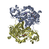
 Movie
Movie Controller


+ Open data

- Basic information
| Entry | Database: PDB / ID: 1pkl | ||||||
|---|---|---|---|---|---|---|---|
| Title | THE STRUCTURE OF LEISHMANIA PYRUVATE KINASE | ||||||
 Components Components | PROTEIN (PYRUVATE KINASE) | ||||||
 Keywords Keywords | TRANSFERASE / PYRUVATE KINASE / GLYCOLYTIC ENZYME / HOMOTETRAMER | ||||||
| Function / homology |  Function and homology information Function and homology informationpyruvate kinase / pyruvate kinase activity / potassium ion binding / response to stress / kinase activity / magnesium ion binding / ATP binding Similarity search - Function | ||||||
| Biological species |   Leishmania mexicana (eukaryote) Leishmania mexicana (eukaryote) | ||||||
| Method |  X-RAY DIFFRACTION / SYNCHROTRON / OTHER / Resolution: 2.35 Å X-RAY DIFFRACTION / SYNCHROTRON / OTHER / Resolution: 2.35 Å | ||||||
 Authors Authors | Rigden, D.J. / Phillips, S.E.V. / Michels, P.A.M. / Fothergill-Gilmore, L.A. | ||||||
 Citation Citation | Journal: J.Mol.Biol. / Year: 1999 Title: The structure of pyruvate kinase from Leishmania mexicana reveals details of the allosteric transition and unusual effector specificity. Authors: Rigden, D.J. / Phillips, S.E. / Michels, P.A. / Fothergill-Gilmore, L.A. | ||||||
| History |
|
- Structure visualization
| Structure viewer | Molecule: MolmilJmol/JSmol |
|---|
- Downloads & links
-Download
| PDBx/mmCIF format | 1pkl.cif.gz | 736.2 KB | Display | PDBx/mmCIF format |
|---|---|---|---|---|
| PDB format | pdb1pkl.ent.gz | 613.7 KB | Display | PDB format |
| PDBx/mmJSON format | 1pkl.json.gz | Tree view | PDBx/mmJSON format | |
| Others |  Other downloads Other downloads |
-Validation report
| Arichive directory | https://data.pdbj.org/pub/pdb/validation_reports/pk/1pklftp://data.pdbj.org/pub/pdb/validation_reports/pk/1pkl | HTTPS FTP |
|---|
-Related structure data
| Related structure data |  1pkyS S: Starting model for refinement |
|---|---|
| Similar structure data |

-Links
 PDBj
PDBj
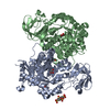
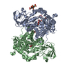
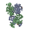
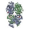
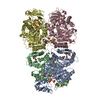
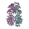
- Assembly
| Deposited unit | 
| ||||||||
|---|---|---|---|---|---|---|---|---|---|
| 1 | 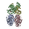
| ||||||||
| 2 | 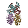
| ||||||||
| Unit cell |
|
-Components
| #1: Protein | Mass: 54467.934 Da / Num. of mol.: 8 Source method: isolated from a genetically manipulated source Source: (gene. exp.) Leishmania mexicana (eukaryote) / Strain: MHOM/BZ/84/BEL46 / Cellular location: CYTOSOL / Gene: PYK / Plasmid: PET3A / Cellular location (production host): CYTOSOL / Gene (production host): PYK / Production host:  #2: Chemical | ChemComp-SO4 /   Mass: 96.063 Da / Num. of mol.: 8 / Source method: obtained synthetically / Formula: SO4 Mass: 96.063 Da / Num. of mol.: 8 / Source method: obtained synthetically / Formula: SO4#3: Water | ChemComp-HOH / |  Mass: 18.015 Da / Num. of mol.: 2952 / Source method: isolated from a natural source / Formula: H2O Mass: 18.015 Da / Num. of mol.: 2952 / Source method: isolated from a natural source / Formula: H2O |
|---|
-Experimental details
-Experiment
| Experiment | Method: X-RAY DIFFRACTION / Number of used crystals: 1 |
|---|
- Sample preparation
| Crystal | Density Matthews: 3.33 Å3/Da / Density % sol: 62.8 % | ||||||||||||||||||||||||||||||
|---|---|---|---|---|---|---|---|---|---|---|---|---|---|---|---|---|---|---|---|---|---|---|---|---|---|---|---|---|---|---|---|
| Crystal grow | pH: 4.8 Details: 6% PEG 4000, 20MM AMMONIUM SULPHATE, 15% GLYCEROL, 20MM SODIUM ACETATE PH 4.8 | ||||||||||||||||||||||||||||||
| Crystal | *PLUS | ||||||||||||||||||||||||||||||
| Crystal grow | *PLUS pH: 4.6 / Method: vapor diffusion, sitting drop | ||||||||||||||||||||||||||||||
| Components of the solutions | *PLUS
|
-Data collection
| Diffraction | Mean temperature: 100 K |
|---|---|
| Diffraction source | Source: SYNCHROTRON / Site: SRS  / Beamline: PX7.2 / Wavelength: 1.488 / Beamline: PX7.2 / Wavelength: 1.488 |
| Detector | Type: MARRESEARCH / Detector: IMAGE PLATE / Date: Dec 15, 1997 / Details: BENT FUSED QUARTZ SINGLE SEGMENT MIRROR |
| Radiation | Monochromator: 200MM LONG BENT, TRIANGULAR GE(111) CRYSTAL / Protocol: SINGLE WAVELENGTH / Monochromatic (M) / Laue (L): M / Scattering type: x-ray |
| Radiation wavelength | Wavelength: 1.488 Å / Relative weight: 1 |
| Reflection | Resolution: 2.35→30 Å / Num. obs: 160789 / % possible obs: 68 % / Redundancy: 1.5 % / Rmerge(I) obs: 0.089 / Net I/σ(I): 5.4 |
| Reflection shell | Resolution: 2.35→2.48 Å / Redundancy: 1.2 % / Rmerge(I) obs: 0.308 / Mean I/σ(I) obs: 2.4 / % possible all: 47.4 |
| Reflection | *PLUS % possible obs: 68 % |
| Reflection shell | *PLUS % possible obs: 47.4 % |
- Processing
| Software |
| ||||||||||||||||||||||||||||||||||||||||||||||||||||||||||||
|---|---|---|---|---|---|---|---|---|---|---|---|---|---|---|---|---|---|---|---|---|---|---|---|---|---|---|---|---|---|---|---|---|---|---|---|---|---|---|---|---|---|---|---|---|---|---|---|---|---|---|---|---|---|---|---|---|---|---|---|---|---|
| Refinement | Method to determine structure: OTHER Starting model: PDB ENTRY 1PKY Resolution: 2.35→30 Å / Rfactor Rfree error: 0.0028 / Data cutoff high absF: 100000 / Data cutoff low absF: 0.001 / Cross valid method: THROUGHOUT / σ(F): 0
| ||||||||||||||||||||||||||||||||||||||||||||||||||||||||||||
| Displacement parameters | Biso mean: 48.9 Å2
| ||||||||||||||||||||||||||||||||||||||||||||||||||||||||||||
| Refinement step | Cycle: LAST / Resolution: 2.35→30 Å
| ||||||||||||||||||||||||||||||||||||||||||||||||||||||||||||
| Refine LS restraints |
| ||||||||||||||||||||||||||||||||||||||||||||||||||||||||||||
| Refine LS restraints NCS | NCS model details: RESTRAINTS | ||||||||||||||||||||||||||||||||||||||||||||||||||||||||||||
| LS refinement shell | Resolution: 2.35→2.43 Å / Rfactor Rfree error: 0.18 / Total num. of bins used: 10
| ||||||||||||||||||||||||||||||||||||||||||||||||||||||||||||
| Xplor file |
| ||||||||||||||||||||||||||||||||||||||||||||||||||||||||||||
| Software | *PLUS Name: X-PLOR / Version: 3.851 / Classification: refinement | ||||||||||||||||||||||||||||||||||||||||||||||||||||||||||||
| Refinement | *PLUS Lowest resolution: 30 Å / σ(F): 0 / % reflection Rfree: 5 % | ||||||||||||||||||||||||||||||||||||||||||||||||||||||||||||
| Solvent computation | *PLUS | ||||||||||||||||||||||||||||||||||||||||||||||||||||||||||||
| Displacement parameters | *PLUS Biso mean: 48.9 Å2 | ||||||||||||||||||||||||||||||||||||||||||||||||||||||||||||
| Refine LS restraints | *PLUS
| ||||||||||||||||||||||||||||||||||||||||||||||||||||||||||||
| LS refinement shell | *PLUS Rfactor Rfree: 0.404 / % reflection Rfree: 5 % / Rfactor Rwork: 0.367 / Rfactor obs: 0.369 |
