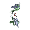
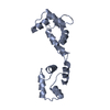
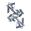
- PDB-3qrx: Chlamydomonas reinhardtii centrin bound to melittin -
+
Open data
ID or keywords:
Loading...
-
Basic information
Entry
Database: PDB / ID: 3qrx
Title
Chlamydomonas reinhardtii centrin bound to melittin
Components
Centrin
Melittin
Keywords
METAL BINDING PROTEIN/TOXIN / calcium-binding / EF-hand / cell division / calcium binding / METAL BINDING PROTEIN-TOXIN complex
Function / homology
Function and homology information
inner dynein arm / dynein heavy chain binding / other organism cell membrane / molecular function inhibitor activity / porin activity / pore complex / protein kinase inhibitor activity / monoatomic ion transport / centriole / microtubule cytoskeleton organization ...inner dynein arm / dynein heavy chain binding / other organism cell membrane / molecular function inhibitor activity / porin activity / pore complex / protein kinase inhibitor activity / monoatomic ion transport / centriole / microtubule cytoskeleton organization / toxin activity / killing of cells of another organism / cell division / calcium ion binding / lipid binding / extracellular region / nucleus Similarity search - Function
Mass: 18.015 Da / Num. of mol.: 31 / Source method: isolated from a natural source / Formula: H2O
-
Experimental details
-
Experiment
Experiment
Method: X-RAY DIFFRACTION / Number of used crystals: 1
-
Sample preparation
Crystal
Density Matthews: 2.32 Å3/Da / Density % sol: 47.08 %
Crystal grow
Temperature: 293 K / Method: vapor diffusion, hanging drop / pH: 7.5 Details: 2 L of the Crcen-MLT (1:1.5 molar ratio, 10mg/mL total) solution with 2 L of a precipitant solution containing 50 mM HEPES, pH 7.5, 0.2 M KCl, and 40% v/v pentaerythritol propoxylate (5/4 ...Details: 2 L of the Crcen-MLT (1:1.5 molar ratio, 10mg/mL total) solution with 2 L of a precipitant solution containing 50 mM HEPES, pH 7.5, 0.2 M KCl, and 40% v/v pentaerythritol propoxylate (5/4 PO/OH), VAPOR DIFFUSION, HANGING DROP, temperature 293K
Resolution: 2.2→20 Å / Cor.coef. Fo:Fc: 0.91 / Cor.coef. Fo:Fc free: 0.885 / SU B: 8.828 / SU ML: 0.228 / Cross valid method: THROUGHOUT / ESU R Free: 0.276 / Stereochemistry target values: MAXIMUM LIKELIHOOD / Details: HYDROGENS HAVE BEEN ADDED IN THE RIDING POSITIONS
Rfactor
Num. reflection
% reflection
Selection details
Rfree
0.34074
522
4.8 %
RANDOM
Rwork
0.29124
-
-
-
all
0.29364
10620
-
-
obs
0.29364
10334
97.31 %
-
Solvent computation
Ion probe radii: 0.8 Å / Shrinkage radii: 0.8 Å / VDW probe radii: 1.4 Å / Solvent model: MASK
Displacement parameters
Biso mean: 40.754 Å2
Baniso -1
Baniso -2
Baniso -3
1-
-1.07 Å2
0 Å2
0 Å2
2-
-
-1.75 Å2
-0 Å2
3-
-
-
2.82 Å2
Refinement step
Cycle: LAST / Resolution: 2.2→20 Å
Protein
Nucleic acid
Ligand
Solvent
Total
Num. atoms
1293
0
4
31
1328
Refine LS restraints
Refine-ID
Type
Dev ideal
Dev ideal target
Number
X-RAY DIFFRACTION
r_bond_refined_d
0.022
0.022
1303
X-RAY DIFFRACTION
r_bond_other_d
0.001
0.02
900
X-RAY DIFFRACTION
r_angle_refined_deg
1.753
1.987
1745
X-RAY DIFFRACTION
r_angle_other_deg
1.004
3
2206
X-RAY DIFFRACTION
r_dihedral_angle_1_deg
6.373
5
163
X-RAY DIFFRACTION
r_dihedral_angle_2_deg
31.648
25.846
65
X-RAY DIFFRACTION
r_dihedral_angle_3_deg
19.751
15
260
X-RAY DIFFRACTION
r_dihedral_angle_4_deg
17.062
15
9
X-RAY DIFFRACTION
r_chiral_restr
0.099
0.2
200
X-RAY DIFFRACTION
r_gen_planes_refined
0.007
0.02
1442
X-RAY DIFFRACTION
r_gen_planes_other
0.001
0.02
242
X-RAY DIFFRACTION
r_mcbond_it
1.095
1.5
814
X-RAY DIFFRACTION
r_mcbond_other
0.238
1.5
337
X-RAY DIFFRACTION
r_mcangle_it
1.978
2
1302
X-RAY DIFFRACTION
r_scbond_it
2.938
3
489
X-RAY DIFFRACTION
r_scangle_it
5.084
4.5
443
LS refinement shell
Resolution: 2.2→2.257 Å / Total num. of bins used: 20
Rfactor
Num. reflection
% reflection
Rfree
0.397
34
-
Rwork
0.342
728
-
obs
-
-
95.37 %
+
About Yorodumi
-
News
-
Feb 9, 2022. New format data for meta-information of EMDB entries
New format data for meta-information of EMDB entries
Version 3 of the EMDB header file is now the official format.
The previous official version 1.9 will be removed from the archive.
In the structure databanks used in Yorodumi, some data are registered as the other names, "COVID-19 virus" and "2019-nCoV". Here are the details of the virus and the list of structure data.
Jan 31, 2019. EMDB accession codes are about to change! (news from PDBe EMDB page)
EMDB accession codes are about to change! (news from PDBe EMDB page)
The allocation of 4 digits for EMDB accession codes will soon come to an end. Whilst these codes will remain in use, new EMDB accession codes will include an additional digit and will expand incrementally as the available range of codes is exhausted. The current 4-digit format prefixed with “EMD-” (i.e. EMD-XXXX) will advance to a 5-digit format (i.e. EMD-XXXXX), and so on. It is currently estimated that the 4-digit codes will be depleted around Spring 2019, at which point the 5-digit format will come into force.
The EM Navigator/Yorodumi systems omit the EMD- prefix.
Related info.:Q: What is EMD? / ID/Accession-code notation in Yorodumi/EM Navigator
Yorodumi is a browser for structure data from EMDB, PDB, SASBDB, etc.
This page is also the successor to EM Navigator detail page, and also detail information page/front-end page for Omokage search.
The word "yorodu" (or yorozu) is an old Japanese word meaning "ten thousand". "mi" (miru) is to see.
Related info.:EMDB / PDB / SASBDB / Comparison of 3 databanks / Yorodumi Search / Aug 31, 2016. New EM Navigator & Yorodumi / Yorodumi Papers / Jmol/JSmol / Function and homology information / Changes in new EM Navigator and Yorodumi
 Movie
Movie Controller


 Open data
 Basic information
 Components
 Keywords
 Function and homology information
 Chlamydomonas reinhardtii (plant)
 X-RAY DIFFRACTION /
  Authors
 Citation
 Structure visualization
 Downloads & links
 Other downloads
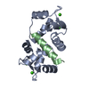


 PDBj
PDBj Assembly
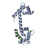


 Mass: 40.078 Da / Num. of mol.: 4 / Source method: obtained synthetically / Formula: Ca
 Mass: 18.015 Da / Num. of mol.: 31 / Source method: isolated from a natural source / Formula: H2O
 Sample preparation
 Processing