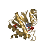
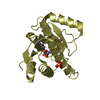
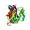
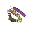
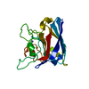
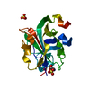
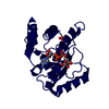
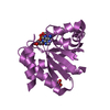
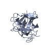
- PDB-3qr5: Structure of the first domain of a cardiac Ryanodine Receptor mut... -
+
データを開く
IDまたはキーワード:
読み込み中...
-
基本情報
登録情報
データベース: PDB / ID: 3qr5
タイトル
Structure of the first domain of a cardiac Ryanodine Receptor mutant with exon 3 deleted
要素
Cardiac Ca2+ release channel
キーワード
SIGNALING PROTEIN / beta trefoil / Sarcoplasmic reticulum
機能・相同性
機能・相同性情報
establishment of protein localization to endoplasmic reticulum / type B pancreatic cell apoptotic process / Purkinje myocyte to ventricular cardiac muscle cell signaling / regulation of atrial cardiac muscle cell action potential / left ventricular cardiac muscle tissue morphogenesis / suramin binding / regulation of AV node cell action potential / regulation of SA node cell action potential / Stimuli-sensing channels / regulation of ventricular cardiac muscle cell action potential ...establishment of protein localization to endoplasmic reticulum / type B pancreatic cell apoptotic process / Purkinje myocyte to ventricular cardiac muscle cell signaling / regulation of atrial cardiac muscle cell action potential / left ventricular cardiac muscle tissue morphogenesis / suramin binding / regulation of AV node cell action potential / regulation of SA node cell action potential / Stimuli-sensing channels / regulation of ventricular cardiac muscle cell action potential / ventricular cardiac muscle cell action potential / positive regulation of sequestering of calcium ion / Ion homeostasis / embryonic heart tube morphogenesis / cardiac muscle hypertrophy / calcium ion transport into cytosol / ryanodine-sensitive calcium-release channel activity / response to caffeine / release of sequestered calcium ion into cytosol by sarcoplasmic reticulum / response to redox state / cellular response to caffeine / calcium ion transmembrane import into cytosol / response to muscle activity / protein kinase A regulatory subunit binding / protein kinase A catalytic subunit binding / positive regulation of the force of heart contraction / smooth endoplasmic reticulum / intracellularly gated calcium channel activity / detection of calcium ion / regulation of cardiac muscle contraction by regulation of the release of sequestered calcium ion / positive regulation of heart rate / response to muscle stretch / cellular response to epinephrine stimulus / calcium channel complex / sarcoplasmic reticulum membrane / regulation of heart rate / sarcoplasmic reticulum / sarcomere / establishment of localization in cell / calcium-mediated signaling / calcium ion transmembrane transport / calcium channel activity / Z disc / intracellular calcium ion homeostasis / calcium ion transport / calmodulin binding / response to hypoxia / calcium ion binding / protein kinase binding / enzyme binding / protein-containing complex / identical protein binding / membrane 類似検索 - 分子機能
 ムービー
ムービー コントローラー


 データを開く
 基本情報
基本情報 要素
要素 キーワード
キーワード 機能・相同性情報
 X線回折 /
  データ登録者
 引用
引用 構造の表示
構造の表示 ダウンロードとリンク
 その他のダウンロード


 PDBj
PDBj


 集合体
集合体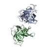


 分子量: 18.015 Da / 分子数: 42 / 由来タイプ: 天然 / 式: H2O
 試料調製
試料調製 / ビームライン: 08ID-1 / 波長: 0.9729 Å
 解析
解析