 Movie
Movie Controller


+ Open data

- Basic information
| Entry | Database: PDB / ID: 1eso | ||||||
|---|---|---|---|---|---|---|---|
| Title | MONOMERIC CU,ZN SUPEROXIDE DISMUTASE FROM ESCHERICHIA COLI | ||||||
 Components Components | CU, ZN SUPEROXIDE DISMUTASE | ||||||
 Keywords Keywords | OXIDOREDUCTASE / CU / ZN SUPEROXIDE DISMUTASE / MONOMERIC SUPEROXIDE DISMUTASE / COPPER ENZYMES / ENZYME EVOLUTION / X-RAY CRYSTAL STRUCTURE | ||||||
| Function / homology |  Function and homology information Function and homology informationsuperoxide metabolic process / superoxide dismutase / superoxide dismutase activity / removal of superoxide radicals / outer membrane-bounded periplasmic space / periplasmic space / copper ion binding / zinc ion binding / metal ion binding Similarity search - Function | ||||||
| Biological species |  | ||||||
| Method |  X-RAY DIFFRACTION / MOLECULAR REPLACEMENT / Resolution: 2 Å X-RAY DIFFRACTION / MOLECULAR REPLACEMENT / Resolution: 2 Å | ||||||
 Authors Authors | Pesce, A. / Capasso, C. / Battistoni, A. / Folcarelli, S. / Rotilio, G. / Desideri, A. / Bolognesi, M. | ||||||
 Citation Citation | Journal: J.Mol.Biol. / Year: 1997 Title: Unique structural features of the monomeric Cu,Zn superoxide dismutase from Escherichia coli, revealed by X-ray crystallography. Authors: Pesce, A. / Capasso, C. / Battistoni, A. / Folcarelli, S. / Rotilio, G. / Desideri, A. / Bolognesi, M. | ||||||
| History |
|
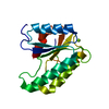
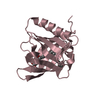
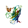
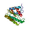
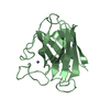
- Structure visualization
| Structure viewer | Molecule: MolmilJmol/JSmol |
|---|
- Downloads & links
-Download
| PDBx/mmCIF format | 1eso.cif.gz | 43.4 KB | Display | PDBx/mmCIF format |
|---|---|---|---|---|
| PDB format | pdb1eso.ent.gz | 28.7 KB | Display | PDB format |
| PDBx/mmJSON format | 1eso.json.gz | Tree view | PDBx/mmJSON format | |
| Others |  Other downloads Other downloads |
-Validation report
| Arichive directory | https://data.pdbj.org/pub/pdb/validation_reports/es/1esoftp://data.pdbj.org/pub/pdb/validation_reports/es/1eso | HTTPS FTP |
|---|
-Related structure data
| Similar structure data |
|---|


-Links
 PDBj
PDBj
- Assembly
| Deposited unit | 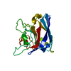
| ||||||||
|---|---|---|---|---|---|---|---|---|---|
| 1 |
| ||||||||
| Unit cell |
|
-Components
| #1: Protein | Mass: 15760.583 Da / Num. of mol.: 1 Source method: isolated from a genetically manipulated source Source: (gene. exp.) References: UniProt: P53635, UniProt: P0AGD1*PLUS, superoxide dismutase |
|---|---|
| #2: Chemical | ChemComp-ZN /   Mass: 65.409 Da / Num. of mol.: 1 / Source method: obtained synthetically / Formula: Zn Mass: 65.409 Da / Num. of mol.: 1 / Source method: obtained synthetically / Formula: Zn |
| #3: Chemical | ChemComp-CU /   Mass: 63.546 Da / Num. of mol.: 1 / Source method: obtained synthetically / Formula: Cu Mass: 63.546 Da / Num. of mol.: 1 / Source method: obtained synthetically / Formula: Cu |
| #4: Water | ChemComp-HOH /  Mass: 18.015 Da / Num. of mol.: 75 / Source method: isolated from a natural source / Formula: H2O Mass: 18.015 Da / Num. of mol.: 75 / Source method: isolated from a natural source / Formula: H2O |
| Has protein modification | Y |
-Experimental details
-Experiment
| Experiment | Method: X-RAY DIFFRACTION / Number of used crystals: 1 |
|---|
- Sample preparation
| Crystal | Density Matthews: 2.1 Å3/Da / Density % sol: 40 % | |||||||||||||||||||||||||||||||||||
|---|---|---|---|---|---|---|---|---|---|---|---|---|---|---|---|---|---|---|---|---|---|---|---|---|---|---|---|---|---|---|---|---|---|---|---|---|
| Crystal grow | pH: 8.5 Details: PROTEIN WAS CRYSTALLIZED FROM 30% PEG 4000, 0.2 M MGCL2, 0.1 M TRIS, PH 8.5 | |||||||||||||||||||||||||||||||||||
| Crystal | *PLUS Density % sol: 40 % | |||||||||||||||||||||||||||||||||||
| Crystal grow | *PLUS Method: vapor diffusion | |||||||||||||||||||||||||||||||||||
| Components of the solutions | *PLUS
|
-Data collection
| Diffraction | Mean temperature: 295 K |
|---|---|
| Diffraction source | Source: ROTATING ANODE / Type: RIGAKU RUH2R / Wavelength: 1.5418 |
| Detector | Type: RIGAKU RAXIS IIC / Detector: IMAGE PLATE / Date: Apr 7, 1996 / Details: PINHOLE COLLIMATOR |
| Radiation | Monochromator: GRAPHITE(002) / Monochromatic (M) / Laue (L): M / Scattering type: x-ray |
| Radiation wavelength | Wavelength: 1.5418 Å / Relative weight: 1 |
| Reflection | Resolution: 2→26.3 Å / Num. obs: 9044 / % possible obs: 96.4 % / Observed criterion σ(I): 0 / Redundancy: 3.1 % / Rmerge(I) obs: 0.06 / Rsym value: 0.06 / Net I/σ(I): 17 |
| Reflection shell | Resolution: 2→2.1 Å / Redundancy: 2.8 % / Rmerge(I) obs: 0.18 / Mean I/σ(I) obs: 3.5 / Rsym value: 0.07 / % possible all: 87 |
| Reflection | *PLUS Num. measured all: 28077 |
- Processing
| Software |
| ||||||||||||||||||||||||||||||||||||||||||||||||||
|---|---|---|---|---|---|---|---|---|---|---|---|---|---|---|---|---|---|---|---|---|---|---|---|---|---|---|---|---|---|---|---|---|---|---|---|---|---|---|---|---|---|---|---|---|---|---|---|---|---|---|---|
| Refinement | Method to determine structure: MOLECULAR REPLACEMENT Starting model: SUPEROXIDE DISMUTASE FROM P.LEIOGNATHI, SOLVED BY SIRAS IN THE DEPOSITORS' LAB Resolution: 2→26 Å / Isotropic thermal model: TNT BCORREL / σ(F): 0 / Stereochemistry target values: TNT: ENGH-HUBER
| ||||||||||||||||||||||||||||||||||||||||||||||||||
| Solvent computation | Solvent model: TNT / Bsol: 103.3 Å2 / ksol: 0.76 e/Å3 | ||||||||||||||||||||||||||||||||||||||||||||||||||
| Refinement step | Cycle: LAST / Resolution: 2→26 Å
| ||||||||||||||||||||||||||||||||||||||||||||||||||
| Refine LS restraints |
| ||||||||||||||||||||||||||||||||||||||||||||||||||
| Software | *PLUS Name: TNT / Version: 5B / Classification: refinement | ||||||||||||||||||||||||||||||||||||||||||||||||||
| Refinement | *PLUS Num. reflection all: 9057 / Num. reflection obs: 8624 / Rfactor all: 0.168 / Rfactor Rfree: 0.263 | ||||||||||||||||||||||||||||||||||||||||||||||||||
| Solvent computation | *PLUS | ||||||||||||||||||||||||||||||||||||||||||||||||||
| Displacement parameters | *PLUS | ||||||||||||||||||||||||||||||||||||||||||||||||||
| Refine LS restraints | *PLUS
|
