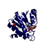
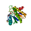
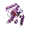
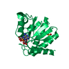
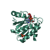
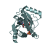
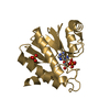
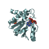
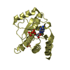
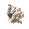
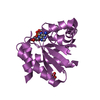
O-acetyl-ADP-ribosedeacetylase / Regulator of RNase III activity
Mass: 19728.211 Da / Num. of mol.: 1 Source method: isolated from a genetically manipulated source Source: (gene. exp.) Escherichia coli K12 (bacteria) / Strain: K12 / Gene: ymdB / Production host: Escherichia coli (E. coli) References: UniProt: P0A8D6, Hydrolases; Acting on carbon-nitrogen bonds, other than peptide bonds; In linear amides
Resolution: 1.8→29.04 Å / Cor.coef. Fo:Fc: 0.959 / Cor.coef. Fo:Fc free: 0.943 / SU B: 2.666 / SU ML: 0.084 / Cross valid method: THROUGHOUT / ESU R: 0.135 / ESU R Free: 0.123 / Stereochemistry target values: MAXIMUM LIKELIHOOD / Details: HYDROGENS HAVE BEEN ADDED IN THE RIDING POSITIONS
Rfactor
Num. reflection
% reflection
Selection details
Rfree
0.21726
791
5 %
RANDOM
Rwork
0.1878
-
-
-
obs
0.1893
15038
98.88 %
-
Solvent computation
Ion probe radii: 0.8 Å / Shrinkage radii: 0.8 Å / VDW probe radii: 1.2 Å / Solvent model: MASK
 Movie
Movie Controller


 Open data
 Basic information
 Components
 Keywords
 Function and homology information
 X-RAY DIFFRACTION /
  Authors
 Citation
 Structure visualization
 Downloads & links
 Other downloads
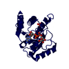

 PDBj
PDBj
 Assembly

 Mass: 559.316 Da / Num. of mol.: 1 / Source method: obtained synthetically / Formula: C15H23N5O14P2
 Mass: 18.015 Da / Num. of mol.: 137 / Source method: isolated from a natural source / Formula: H2O
 Sample preparation
 / Beamline: BL17U / Wavelength: 0.9791 Å
 Processing