Type: MARMOSAIC 300 mm CCD / Detector: CCD / Date: Mar 10, 2010
Radiation
Monochromator: double crystal monochromator and K-B pair of biomorph mirrors for vertical and horizontal focusing Protocol: SINGLE WAVELENGTH / Monochromatic (M) / Laue (L): M / Scattering type: x-ray
Radiation wavelength
Wavelength: 1.033 Å / Relative weight: 1
Reflection
Resolution: 1.6→50 Å / Num. all: 35695 / Num. obs: 35547 / % possible obs: 99.6 %
Reflection shell
Resolution: 1.6→1.642 Å / % possible all: 97.6
-
Processing
Software
Name
Version
Classification
Blu-Ice
datacollection
PHASER
phasing
REFMAC
5.5.0109
refinement
HKL-2000
datareduction
XPREP
datareduction
Refinement
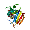
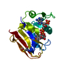
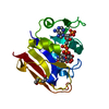
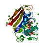
Method to determine structure: MOLECULAR REPLACEMENT / Resolution: 1.6→49.63 Å / Cor.coef. Fo:Fc: 0.947 / Cor.coef. Fo:Fc free: 0.922 / SU B: 3.663 / SU ML: 0.07 / Cross valid method: THROUGHOUT / ESU R Free: 0.093 / Stereochemistry target values: MAXIMUM LIKELIHOOD / Details: HYDROGENS HAVE BEEN ADDED IN THE RIDING POSITIONS
Rfactor
Num. reflection
% reflection
Selection details
Rfree
0.24534
1639
5 %
RANDOM
Rwork
0.20849
-
-
-
obs
0.21029
31133
93.71 %
-
Solvent computation
Ion probe radii: 0.8 Å / Shrinkage radii: 0.8 Å / VDW probe radii: 1.4 Å / Solvent model: MASK
Displacement parameters
Biso mean: 14.373 Å2
Baniso -1
Baniso -2
Baniso -3
1-
0.28 Å2
0.14 Å2
-0 Å2
2-
-
0.28 Å2
-0 Å2
3-
-
-
-0.42 Å2
Refinement step
Cycle: LAST / Resolution: 1.6→49.63 Å
Protein
Nucleic acid
Ligand
Solvent
Total
Num. atoms
1273
0
83
199
1555
Refine LS restraints
Refine-ID
Type
Dev ideal
Dev ideal target
Number
X-RAY DIFFRACTION
r_bond_refined_d
0.011
0.022
1457
X-RAY DIFFRACTION
r_bond_other_d
0.002
0.02
970
X-RAY DIFFRACTION
r_angle_refined_deg
1.539
2.028
1999
X-RAY DIFFRACTION
r_angle_other_deg
0.886
3
2361
X-RAY DIFFRACTION
r_dihedral_angle_1_deg
5.334
5
168
X-RAY DIFFRACTION
r_dihedral_angle_2_deg
35.153
23.75
64
X-RAY DIFFRACTION
r_dihedral_angle_3_deg
13.266
15
224
X-RAY DIFFRACTION
r_dihedral_angle_4_deg
14.257
15
10
X-RAY DIFFRACTION
r_chiral_restr
0.086
0.2
208
X-RAY DIFFRACTION
r_gen_planes_refined
0.007
0.021
1596
X-RAY DIFFRACTION
r_gen_planes_other
0.002
0.02
277
X-RAY DIFFRACTION
r_mcbond_it
0.589
1.5
844
X-RAY DIFFRACTION
r_mcbond_other
0.17
1.5
331
X-RAY DIFFRACTION
r_mcangle_it
1.075
2
1376
X-RAY DIFFRACTION
r_scbond_it
1.889
3
613
X-RAY DIFFRACTION
r_scangle_it
3.082
4.5
622
LS refinement shell
Resolution: 1.6→1.642 Å / Total num. of bins used: 20
Rfactor
Num. reflection
% reflection
Rfree
0.317
115
-
Rwork
0.293
2320
-
obs
-
-
95.9 %
Refinement TLS params.
Method: refined / Refine-ID: X-RAY DIFFRACTION
ID
L11 (°2)
L12 (°2)
L13 (°2)
L22 (°2)
L23 (°2)
L33 (°2)
S11 (Å °)
S12 (Å °)
S13 (Å °)
S21 (Å °)
S22 (Å °)
S23 (Å °)
S31 (Å °)
S32 (Å °)
S33 (Å °)
T11 (Å2)
T12 (Å2)
T13 (Å2)
T22 (Å2)
T23 (Å2)
T33 (Å2)
Origin x (Å)
Origin y (Å)
Origin z (Å)
1
3.2157
-0.3416
-0.9324
0.9694
0.1513
1.4604
0.0474
0.1176
0.0281
-0.0482
-0.0109
-0.0091
0.0278
-0.0116
-0.0365
0.02
-0.0047
0.0032
0.0262
0.0018
0.0022
-2.505
36.2578
5.5608
2
4.6111
-0.8265
-0.3121
0.7455
-0.2286
2.626
-0.0268
0.2691
-0.4275
-0.0065
0.0344
0.1672
0.2903
-0.1558
-0.0077
0.0508
-0.0465
0.002
0.0912
-0.0168
0.057
-16.2558
30.7826
8.828
Refinement TLS group
ID
Refine-ID
Refine TLS-ID
Auth asym-ID
Auth seq-ID
1
X-RAY DIFFRACTION
1
A
1 - 102
2
X-RAY DIFFRACTION
2
A
103 - 160
+
About Yorodumi
-
News
-
Feb 9, 2022. New format data for meta-information of EMDB entries
New format data for meta-information of EMDB entries
Version 3 of the EMDB header file is now the official format.
The previous official version 1.9 will be removed from the archive.
In the structure databanks used in Yorodumi, some data are registered as the other names, "COVID-19 virus" and "2019-nCoV". Here are the details of the virus and the list of structure data.
Jan 31, 2019. EMDB accession codes are about to change! (news from PDBe EMDB page)
EMDB accession codes are about to change! (news from PDBe EMDB page)
The allocation of 4 digits for EMDB accession codes will soon come to an end. Whilst these codes will remain in use, new EMDB accession codes will include an additional digit and will expand incrementally as the available range of codes is exhausted. The current 4-digit format prefixed with “EMD-” (i.e. EMD-XXXX) will advance to a 5-digit format (i.e. EMD-XXXXX), and so on. It is currently estimated that the 4-digit codes will be depleted around Spring 2019, at which point the 5-digit format will come into force.
The EM Navigator/Yorodumi systems omit the EMD- prefix.
Related info.:Q: What is EMD? / ID/Accession-code notation in Yorodumi/EM Navigator
Yorodumi is a browser for structure data from EMDB, PDB, SASBDB, etc.
This page is also the successor to EM Navigator detail page, and also detail information page/front-end page for Omokage search.
The word "yorodu" (or yorozu) is an old Japanese word meaning "ten thousand". "mi" (miru) is to see.
Related info.:EMDB / PDB / SASBDB / Comparison of 3 databanks / Yorodumi Search / Aug 31, 2016. New EM Navigator & Yorodumi / Yorodumi Papers / Jmol/JSmol / Function and homology information / Changes in new EM Navigator and Yorodumi
 Movie
Movie Controller

 Yorodumi
 Open data
 Basic information
 Components
 Keywords
 Function and homology information
 X-RAY DIFFRACTION /
  Authors
 Citation
 Structure visualization
 Downloads & links
 Other downloads


 PDBj
PDBj
 Assembly

 Mass: 441.397 Da / Num. of mol.: 1 / Source method: obtained synthetically / Formula: C19H19N7O6
 Mass: 743.405 Da / Num. of mol.: 1 / Source method: obtained synthetically / Formula: C21H28N7O17P3
 Mass: 22.990 Da / Num. of mol.: 3 / Source method: obtained synthetically / Formula: Na
 Mass: 18.015 Da / Num. of mol.: 199 / Source method: isolated from a natural source / Formula: H2O
 Sample preparation
 / Beamline: 23-ID-D / Wavelength: 1.033 Å
 Processing