| Entry | Database: PDB / ID: 3qkx
|
|---|
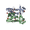
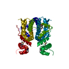
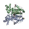
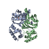
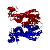
| Title | Crystal structure of a TetR-family transcriptional regulator (HI0893) from Haemophilus influenzae RD at 2.35 A resolution |
|---|
 Components Components | Uncharacterized HTH-type transcriptional regulator HI_0893 |
|---|
 Keywords Keywords | Transcription regulator / STRUCTURAL GENOMICS / JOINT CENTER FOR STRUCTURAL GENOMICS / JCSG / PROTEIN STRUCTURE INITIATIVE / PSI-BIOLOGY / TRANSCRIPTION |
|---|
| Function / homology |  Function and homology information Function and homology information
transcription cis-regulatory region binding / DNA-binding transcription factor activity / regulation of DNA-templated transcriptionSimilarity search - Function : / Tetracyclin repressor-like HI_0893, C-terminal domain / : / DNA-binding HTH domain, TetR-type, conserved site / TetR-type HTH domain signature. / Tetracycline Repressor, domain 2 / Tetracycline Repressor; domain 2 / Bacterial regulatory proteins, tetR family / DNA-binding HTH domain, TetR-type / TetR-type HTH domain profile. ...: / Tetracyclin repressor-like HI_0893, C-terminal domain / : / DNA-binding HTH domain, TetR-type, conserved site / TetR-type HTH domain signature. / Tetracycline Repressor, domain 2 / Tetracycline Repressor; domain 2 / Bacterial regulatory proteins, tetR family / DNA-binding HTH domain, TetR-type / TetR-type HTH domain profile. / Homeobox-like domain superfamily / Orthogonal Bundle / Mainly AlphaSimilarity search - Domain/homology |
|---|
| Biological species |  Haemophilus influenzae (bacteria) Haemophilus influenzae (bacteria) |
|---|
| Method |  X-RAY DIFFRACTION / SYNCHROTRON / MAD / Resolution: 2.35 Å X-RAY DIFFRACTION / SYNCHROTRON / MAD / Resolution: 2.35 Å |
|---|
 Authors Authors | Joint Center for Structural Genomics (JCSG) |
|---|
 Citation Citation | Journal: To be published
Title: Crystal structure of a TetR-family transcriptional regulator (HI0893) from Haemophilus influenzae RD at 2.35 A resolution
Authors: Joint Center for Structural Genomics (JCSG) |
|---|
| History | | Deposition | Feb 1, 2011 | Deposition site: RCSB / Processing site: RCSB |
|---|
| Revision 1.0 | Mar 2, 2011 | Provider: repository / Type: Initial release |
|---|
| Revision 1.1 | Jul 13, 2011 | Group: Version format compliance |
|---|
| Revision 1.2 | Jul 20, 2011 | Group: Structure summary |
|---|
| Revision 1.3 | Oct 25, 2017 | Group: Author supporting evidence / Refinement description / Category: pdbx_struct_assembly_auth_evidence / software / Item: _software.classification / _software.name |
|---|
| Revision 1.4 | Feb 1, 2023 | Group: Database references / Derived calculations
Category: database_2 / struct_conn ...database_2 / struct_conn / struct_ref_seq_dif / struct_site
Item: _database_2.pdbx_DOI / _database_2.pdbx_database_accession ..._database_2.pdbx_DOI / _database_2.pdbx_database_accession / _struct_conn.pdbx_leaving_atom_flag / _struct_ref_seq_dif.details / _struct_site.pdbx_auth_asym_id / _struct_site.pdbx_auth_comp_id / _struct_site.pdbx_auth_seq_id |
|---|
| Revision 1.5 | Nov 27, 2024 | Group: Data collection / Refinement description / Structure summary
Category: chem_comp_atom / chem_comp_bond ...chem_comp_atom / chem_comp_bond / pdbx_entry_details / pdbx_modification_feature / struct_ncs_dom_lim
Item: _pdbx_entry_details.has_protein_modification / _struct_ncs_dom_lim.beg_auth_comp_id ..._pdbx_entry_details.has_protein_modification / _struct_ncs_dom_lim.beg_auth_comp_id / _struct_ncs_dom_lim.beg_label_asym_id / _struct_ncs_dom_lim.beg_label_comp_id / _struct_ncs_dom_lim.beg_label_seq_id / _struct_ncs_dom_lim.end_auth_comp_id / _struct_ncs_dom_lim.end_label_asym_id / _struct_ncs_dom_lim.end_label_comp_id / _struct_ncs_dom_lim.end_label_seq_id |
|---|
|
|---|
 Movie
Movie Controller

 Yorodumi
 Open data
 Basic information
 Structure visualization
 Downloads & links
 Other downloads
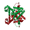


 PDBj
PDBj Assembly