 Movie
Movie Controller

[English] 日本語
 Yorodumi
- PDB-3qkw: Structure of Streptococcus parasangunini Gtf3 glycosyltransferase -
+ Open data

- Basic information
| Entry | Database: PDB / ID: 3qkw | ||||||
|---|---|---|---|---|---|---|---|
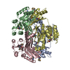
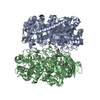
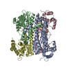
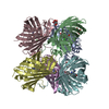
| Title | Structure of Streptococcus parasangunini Gtf3 glycosyltransferase | ||||||
 Components Components | Nucleotide sugar synthetase-like protein | ||||||
 Keywords Keywords | TRANSFERASE / GT-B fold / glycosyltransferase / NUCLEOTIDE SUGAR SYNTHETASE-LIKE PROTEIN | ||||||
| Function / homology |  Function and homology information Function and homology informationUDP-glucosyltransferase activity / glycoprotein biosynthetic process / Transferases; Glycosyltransferases; Hexosyltransferases / nucleotide binding Similarity search - Function | ||||||
| Biological species |  Streptococcus parasanguinis (bacteria) Streptococcus parasanguinis (bacteria) | ||||||
| Method |  X-RAY DIFFRACTION / SYNCHROTRON / MOLECULAR REPLACEMENT / Resolution: 2.287 Å X-RAY DIFFRACTION / SYNCHROTRON / MOLECULAR REPLACEMENT / Resolution: 2.287 Å | ||||||
 Authors Authors | Zhu, F. / Erlandsen, H. / Huang, Y. / Ding, L. / Zhou, M. / Liang, X. / Ma, J.-B. / Wu, H. | ||||||
 Citation Citation | Journal: J.Biol.Chem. / Year: 2011 Title: Structural and Functional Analysis of a New Subfamily of Glycosyltransferases Required for Glycosylation of Serine-rich Streptococcal Adhesins. Authors: Zhu, F. / Erlandsen, H. / Ding, L. / Li, J. / Huang, Y. / Zhou, M. / Liang, X. / Ma, J. / Wu, H. | ||||||
| History |
|
- Structure visualization
| Structure viewer | Molecule: MolmilJmol/JSmol |
|---|
- Downloads & links
-Download
| PDBx/mmCIF format | 3qkw.cif.gz | 530.5 KB | Display | PDBx/mmCIF format |
|---|---|---|---|---|
| PDB format | pdb3qkw.ent.gz | 439.6 KB | Display | PDB format |
| PDBx/mmJSON format | 3qkw.json.gz | Tree view | PDBx/mmJSON format | |
| Others |  Other downloads Other downloads |
-Validation report
| Arichive directory | https://data.pdbj.org/pub/pdb/validation_reports/qk/3qkwftp://data.pdbj.org/pub/pdb/validation_reports/qk/3qkw | HTTPS FTP |
|---|
-Related structure data


-Links
 PDBj
PDBj


- Assembly
| Deposited unit | 
| ||||||||
|---|---|---|---|---|---|---|---|---|---|
| 1 |
| ||||||||
| Unit cell |
|
-Components
| #1: Protein | Mass: 38267.910 Da / Num. of mol.: 4 Source method: isolated from a genetically manipulated source Source: (gene. exp.) Streptococcus parasanguinis (bacteria) / Gene: nss / Plasmid: pET-SUMO-Gtf3 / Production host: #2: Chemical | ChemComp-UDP /   Type: RNA linking / Mass: 404.161 Da / Num. of mol.: 4 / Source method: obtained synthetically / Formula: C9H14N2O12P2 / Comment: UDP*YM Type: RNA linking / Mass: 404.161 Da / Num. of mol.: 4 / Source method: obtained synthetically / Formula: C9H14N2O12P2 / Comment: UDP*YM#3: Water | ChemComp-HOH / |  Mass: 18.015 Da / Num. of mol.: 188 / Source method: isolated from a natural source / Formula: H2O Mass: 18.015 Da / Num. of mol.: 188 / Source method: isolated from a natural source / Formula: H2O |
|---|
-Experimental details
-Experiment
| Experiment | Method: X-RAY DIFFRACTION / Number of used crystals: 1 |
|---|
- Sample preparation
| Crystal | Density Matthews: 2.36 Å3/Da / Density % sol: 47.78 % |
|---|---|
| Crystal grow | Temperature: 293 K / Method: vapor diffusion, hanging drop / pH: 7 Details: 1 ul of protein solution (15 mg/ml protein and 10mM UDP-Glucose) was mixed with 1 ul of reservoir (0.1M Succinic Acid, 11% Polyethylene glycol 3,350, 10% glycerol), pH 7.0, VAPOR DIFFUSION, ...Details: 1 ul of protein solution (15 mg/ml protein and 10mM UDP-Glucose) was mixed with 1 ul of reservoir (0.1M Succinic Acid, 11% Polyethylene glycol 3,350, 10% glycerol), pH 7.0, VAPOR DIFFUSION, HANGING DROP, temperature 293K |
-Data collection
| Diffraction | Mean temperature: 100 K |
|---|---|
| Diffraction source | Source: SYNCHROTRON / Site: APS  / Beamline: 22-ID / Wavelength: 1.033 Å / Beamline: 22-ID / Wavelength: 1.033 Å |
| Detector | Type: MARMOSAIC 300 mm CCD / Detector: CCD / Date: Apr 5, 2010 |
| Radiation | Monochromator: Double crystal - liquid nitrogen cooled / Protocol: SAD / Monochromatic (M) / Laue (L): M / Scattering type: x-ray |
| Radiation wavelength | Wavelength: 1.033 Å / Relative weight: 1 |
| Reflection | Resolution: 2.1→42.512 Å / Num. obs: 63229 / % possible obs: 98.2 % / Observed criterion σ(F): 0 / Observed criterion σ(I): 0 / Redundancy: 5.6 % / Rmerge(I) obs: 0.09 / Net I/σ(I): 16.2 |
| Reflection shell | Resolution: 2.1→2.18 Å / Redundancy: 3.5 % / Rmerge(I) obs: 0.479 / Mean I/σ(I) obs: 10.8 / % possible all: 96 |
- Processing
| Software |
| |||||||||||||||||||||||||||||||||||||||||||||||||||||||||||||||||||||||||||||||||||||||||||||||||||||||||||||||||||||||||||||
|---|---|---|---|---|---|---|---|---|---|---|---|---|---|---|---|---|---|---|---|---|---|---|---|---|---|---|---|---|---|---|---|---|---|---|---|---|---|---|---|---|---|---|---|---|---|---|---|---|---|---|---|---|---|---|---|---|---|---|---|---|---|---|---|---|---|---|---|---|---|---|---|---|---|---|---|---|---|---|---|---|---|---|---|---|---|---|---|---|---|---|---|---|---|---|---|---|---|---|---|---|---|---|---|---|---|---|---|---|---|---|---|---|---|---|---|---|---|---|---|---|---|---|---|---|---|---|
| Refinement | Method to determine structure: MOLECULAR REPLACEMENT Starting model: A SAD dataset was collected using Se-Met crystal. Initial structure determined in spacegroup C2221 with Gtf3 dimer. Resolution: 2.287→42.512 Å / SU ML: 0.37 / Isotropic thermal model: Anisotropic / σ(F): 1.34 / Phase error: 28.16 / Stereochemistry target values: ML
| |||||||||||||||||||||||||||||||||||||||||||||||||||||||||||||||||||||||||||||||||||||||||||||||||||||||||||||||||||||||||||||
| Solvent computation | Shrinkage radii: 0.9 Å / VDW probe radii: 1.11 Å / Solvent model: FLAT BULK SOLVENT MODEL / Bsol: 45.737 Å2 / ksol: 0.338 e/Å3 | |||||||||||||||||||||||||||||||||||||||||||||||||||||||||||||||||||||||||||||||||||||||||||||||||||||||||||||||||||||||||||||
| Displacement parameters |
| |||||||||||||||||||||||||||||||||||||||||||||||||||||||||||||||||||||||||||||||||||||||||||||||||||||||||||||||||||||||||||||
| Refinement step | Cycle: LAST / Resolution: 2.287→42.512 Å
| |||||||||||||||||||||||||||||||||||||||||||||||||||||||||||||||||||||||||||||||||||||||||||||||||||||||||||||||||||||||||||||
| Refine LS restraints |
| |||||||||||||||||||||||||||||||||||||||||||||||||||||||||||||||||||||||||||||||||||||||||||||||||||||||||||||||||||||||||||||
| LS refinement shell |
| |||||||||||||||||||||||||||||||||||||||||||||||||||||||||||||||||||||||||||||||||||||||||||||||||||||||||||||||||||||||||||||
| Refinement TLS params. | Method: refined / Refine-ID: X-RAY DIFFRACTION
| |||||||||||||||||||||||||||||||||||||||||||||||||||||||||||||||||||||||||||||||||||||||||||||||||||||||||||||||||||||||||||||
| Refinement TLS group |
|
