 Movie
Movie Controller

[English] 日本語
 Yorodumi
- PDB-3qhy: Structural, thermodynamic and kinetic analysis of the picomolar b... -
+ Open data

- Basic information
| Entry | Database: PDB / ID: 3qhy | ||||||
|---|---|---|---|---|---|---|---|
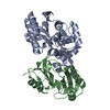
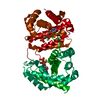
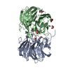
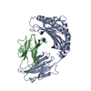
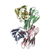
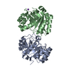
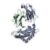
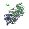
| Title | Structural, thermodynamic and kinetic analysis of the picomolar binding affinity interaction of the beta-lactamase inhibitor protein-II (BLIP-II) with class A beta-lactamases | ||||||
 Components Components |
| ||||||
 Keywords Keywords | HYDROLASE/HYDROLASE INHIBITOR / enyzme-inhibitor complex / beta-propeller / beta-lactamase / protein:protein interaction / HYDROLASE-HYDROLASE INHIBITOR complex | ||||||
| Function / homology |  Function and homology information Function and homology informationbeta-lactam antibiotic catabolic process / beta-lactamase activity / beta-lactamase / ubiquitin protein ligase activity / response to antibiotic / metal ion binding Similarity search - Function | ||||||
| Biological species |  Streptomyces exfoliatus (bacteria) Streptomyces exfoliatus (bacteria) | ||||||
| Method |  X-RAY DIFFRACTION / SYNCHROTRON / MOLECULAR REPLACEMENT / Resolution: 2.06 Å X-RAY DIFFRACTION / SYNCHROTRON / MOLECULAR REPLACEMENT / Resolution: 2.06 Å | ||||||
 Authors Authors | Brown, N.G. / Chow, D.C. / Sankaran, B. / Zwart, P. / Prasad, B.V.V. / Palzkill, T. / Berkeley Structural Genomics Center (BSGC) | ||||||
 Citation Citation | Journal: J.Biol.Chem. / Year: 2011 Title: Analysis of the binding forces driving the tight interactions between beta-lactamase inhibitory protein-II (BLIP-II) and class A beta-lactamases. Authors: Brown, N.G. / Chow, D.C. / Sankaran, B. / Zwart, P. / Prasad, B.V. / Palzkill, T. | ||||||
| History |
|
- Structure visualization
| Structure viewer | Molecule: MolmilJmol/JSmol |
|---|
- Downloads & links
-Download
| PDBx/mmCIF format | 3qhy.cif.gz | 121.9 KB | Display | PDBx/mmCIF format |
|---|---|---|---|---|
| PDB format | pdb3qhy.ent.gz | 92.1 KB | Display | PDB format |
| PDBx/mmJSON format | 3qhy.json.gz | Tree view | PDBx/mmJSON format | |
| Others |  Other downloads Other downloads |
-Validation report
| Arichive directory | https://data.pdbj.org/pub/pdb/validation_reports/qh/3qhyftp://data.pdbj.org/pub/pdb/validation_reports/qh/3qhy | HTTPS FTP |
|---|
-Related structure data
| Related structure data |  3qi0C  1jtdS S: Starting model for refinement C: citing same article ( |
|---|---|
| Similar structure data |


-Links
 PDBj
PDBj


- Assembly
| Deposited unit | 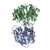
| ||||||||
|---|---|---|---|---|---|---|---|---|---|
| 1 |
| ||||||||
| Unit cell |
|
-Components
| #1: Protein | Mass: 29759.611 Da / Num. of mol.: 1 / Fragment: unp residues 39-309 Source method: isolated from a genetically manipulated source Source: (gene. exp.) |
|---|---|
| #2: Protein | Mass: 28360.947 Da / Num. of mol.: 1 / Fragment: unp residues 41-311 Source method: isolated from a genetically manipulated source Source: (gene. exp.) Streptomyces exfoliatus (bacteria) / Gene: bliB / Production host: |
| #3: Water | ChemComp-HOH /  Mass: 18.015 Da / Num. of mol.: 508 / Source method: isolated from a natural source / Formula: H2O Mass: 18.015 Da / Num. of mol.: 508 / Source method: isolated from a natural source / Formula: H2O |
-Experimental details
-Experiment
| Experiment | Method: X-RAY DIFFRACTION / Number of used crystals: 1 |
|---|
- Sample preparation
| Crystal | Density Matthews: 2.72 Å3/Da / Density % sol: 54.71 % |
|---|---|
| Crystal grow | Temperature: 298 K / pH: 8.5 Details: 0.1M Bicine, 13 % PEG 10,000, pH 8.5, VAPOR DIFFUSION, HANGING DROP, temperature 298K |
-Data collection
| Diffraction | Mean temperature: 100 K |
|---|---|
| Diffraction source | Source: SYNCHROTRON / Site: ALS  / Beamline: 5.0.1 / Wavelength: 0.98 / Beamline: 5.0.1 / Wavelength: 0.98 |
| Detector | Type: ADSC QUANTUM 315r / Detector: CCD / Date: Apr 10, 2010 |
| Radiation | Monochromator: SINGLE CRYSTAL, CYLINDRICALLY BENT SI(220) / Protocol: SINGLE WAVELENGTH / Monochromatic (M) / Laue (L): M / Scattering type: x-ray |
| Radiation wavelength | Wavelength: 0.98 Å / Relative weight: 1 |
| Reflection | Resolution: 2.06→50 Å / Num. obs: 37880 / % possible obs: 98.4 % / Observed criterion σ(I): -3 |
| Reflection shell | Resolution: 2.06→2.1 Å / % possible all: 91.3 |
- Processing
| Software |
| |||||||||||||||||||||||||||||||||||||||||||||||||||||||||||||||||||||||||||||
|---|---|---|---|---|---|---|---|---|---|---|---|---|---|---|---|---|---|---|---|---|---|---|---|---|---|---|---|---|---|---|---|---|---|---|---|---|---|---|---|---|---|---|---|---|---|---|---|---|---|---|---|---|---|---|---|---|---|---|---|---|---|---|---|---|---|---|---|---|---|---|---|---|---|---|---|---|---|---|
| Refinement | Method to determine structure: MOLECULAR REPLACEMENT Starting model: PDB ENTRY 1JTD Resolution: 2.06→49.95 Å / SU ML: 0.26 / σ(F): 1.93 / Phase error: 20.76 / Stereochemistry target values: ML
| |||||||||||||||||||||||||||||||||||||||||||||||||||||||||||||||||||||||||||||
| Solvent computation | Shrinkage radii: 0.9 Å / VDW probe radii: 1.11 Å / Solvent model: FLAT BULK SOLVENT MODEL / Bsol: 41.74 Å2 / ksol: 0.36 e/Å3 | |||||||||||||||||||||||||||||||||||||||||||||||||||||||||||||||||||||||||||||
| Displacement parameters |
| |||||||||||||||||||||||||||||||||||||||||||||||||||||||||||||||||||||||||||||
| Refinement step | Cycle: LAST / Resolution: 2.06→49.95 Å
| |||||||||||||||||||||||||||||||||||||||||||||||||||||||||||||||||||||||||||||
| Refine LS restraints |
| |||||||||||||||||||||||||||||||||||||||||||||||||||||||||||||||||||||||||||||
| LS refinement shell |
|
