 Movie
Movie Controller


+ Open data

- Basic information
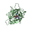
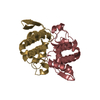
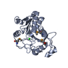
| Entry | Database: PDB / ID: 3qh4 | ||||||
|---|---|---|---|---|---|---|---|
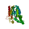
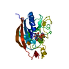
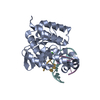
| Title | Crystal structure of esterase LipW from Mycobacterium marinum | ||||||
 Components Components | Esterase LipW | ||||||
 Keywords Keywords | HYDROLASE / Structural Genomics / SSGCID / Seattle Structural Genomics Center for Infectious Disease / Mycobacterium / tuberculosis / marinum / ortholog / LipW / esterase / heroin esterase / multidrug resistance / TB | ||||||
| Function / homology |  Function and homology information Function and homology information | ||||||
| Biological species |  Mycobacterium marinum (bacteria) Mycobacterium marinum (bacteria) | ||||||
| Method |  X-RAY DIFFRACTION / MOLECULAR REPLACEMENT / molecular replacement / Resolution: 1.75 Å X-RAY DIFFRACTION / MOLECULAR REPLACEMENT / molecular replacement / Resolution: 1.75 Å | ||||||
 Authors Authors | Seattle Structural Genomics Center for Infectious Disease (SSGCID) | ||||||
 Citation Citation | Journal: Biochemistry / Year: 2016 Title: Structural Basis for the Strict Substrate Selectivity of the Mycobacterial Hydrolase LipW. Authors: McKary, M.G. / Abendroth, J. / Edwards, T.E. / Johnson, R.J. #1: Journal: Tuberculosis (Edinb) / Year: 2015Title: Increasing the structural coverage of tuberculosis drug targets. Authors: Baugh, L. / Phan, I. / Begley, D.W. / Clifton, M.C. / Armour, B. / Dranow, D.M. / Taylor, B.M. / Muruthi, M.M. / Abendroth, J. / Fairman, J.W. / Fox, D. / Dieterich, S.H. / Staker, B.L. / ...Authors: Baugh, L. / Phan, I. / Begley, D.W. / Clifton, M.C. / Armour, B. / Dranow, D.M. / Taylor, B.M. / Muruthi, M.M. / Abendroth, J. / Fairman, J.W. / Fox, D. / Dieterich, S.H. / Staker, B.L. / Gardberg, A.S. / Choi, R. / Hewitt, S.N. / Napuli, A.J. / Myers, J. / Barrett, L.K. / Zhang, Y. / Ferrell, M. / Mundt, E. / Thompkins, K. / Tran, N. / Lyons-Abbott, S. / Abramov, A. / Sekar, A. / Serbzhinskiy, D. / Lorimer, D. / Buchko, G.W. / Stacy, R. / Stewart, L.J. / Edwards, T.E. / Van Voorhis, W.C. / Myler, P.J. | ||||||
| History |
|
- Structure visualization
| Structure viewer | Molecule: MolmilJmol/JSmol |
|---|
- Downloads & links
-Download
| PDBx/mmCIF format | 3qh4.cif.gz | 136.3 KB | Display | PDBx/mmCIF format |
|---|---|---|---|---|
| PDB format | pdb3qh4.ent.gz | 103.8 KB | Display | PDB format |
| PDBx/mmJSON format | 3qh4.json.gz | Tree view | PDBx/mmJSON format | |
| Others |  Other downloads Other downloads |
-Validation report
| Arichive directory | https://data.pdbj.org/pub/pdb/validation_reports/qh/3qh4ftp://data.pdbj.org/pub/pdb/validation_reports/qh/3qh4 | HTTPS FTP |
|---|
-Related structure data
| Related structure data | 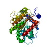 1lzkS S: Starting model for refinement |
|---|---|
| Similar structure data | |
| Other databases |


-Links
 PDBj
PDBj- Assembly
| Deposited unit | 
| ||||||||
|---|---|---|---|---|---|---|---|---|---|
| 1 |
| ||||||||
| Unit cell |
|
-Components
| #1: Protein | Mass: 33837.223 Da / Num. of mol.: 1 Source method: isolated from a genetically manipulated source Source: (gene. exp.) Mycobacterium marinum (bacteria) / Strain: ATCC BAA-535 / M / Gene: lipW, MMAR_0404 / Plasmid: AVA0421 / Production host: |
|---|---|
| #2: Water | ChemComp-HOH /  Mass: 18.015 Da / Num. of mol.: 419 / Source method: isolated from a natural source / Formula: H2O Mass: 18.015 Da / Num. of mol.: 419 / Source method: isolated from a natural source / Formula: H2O |
-Experimental details
-Experiment
| Experiment | Method: X-RAY DIFFRACTION / Number of used crystals: 1 |
|---|
- Sample preparation
| Crystal | Density Matthews: 2.18 Å3/Da / Density % sol: 43.45 % |
|---|---|
| Crystal grow | Temperature: 289 K / Method: vapor diffusion, sitting drop / pH: 8 Details: protein at 55.6 mg/mL, 0.2 M MgCl2, 0.1 M Tris, 20 % PEG 6000, cryo-protected with 25 % ethylene glycol, pH 8.0, VAPOR DIFFUSION, SITTING DROP, temperature 289 K |
-Data collection
| Diffraction | Mean temperature: 100 K | |||||||||||||||||||||||||||||||||||||||||||||||||||||||||||||||||||||||||||||||||||||||||||||||||||||||||||||||||||||||||||||||||||||||||||||||||||||||||||||||||||||||||||||||||||||||||||||
|---|---|---|---|---|---|---|---|---|---|---|---|---|---|---|---|---|---|---|---|---|---|---|---|---|---|---|---|---|---|---|---|---|---|---|---|---|---|---|---|---|---|---|---|---|---|---|---|---|---|---|---|---|---|---|---|---|---|---|---|---|---|---|---|---|---|---|---|---|---|---|---|---|---|---|---|---|---|---|---|---|---|---|---|---|---|---|---|---|---|---|---|---|---|---|---|---|---|---|---|---|---|---|---|---|---|---|---|---|---|---|---|---|---|---|---|---|---|---|---|---|---|---|---|---|---|---|---|---|---|---|---|---|---|---|---|---|---|---|---|---|---|---|---|---|---|---|---|---|---|---|---|---|---|---|---|---|---|---|---|---|---|---|---|---|---|---|---|---|---|---|---|---|---|---|---|---|---|---|---|---|---|---|---|---|---|---|---|---|---|---|
| Diffraction source | Source: ROTATING ANODE / Type: RIGAKU FR-E+ SUPERBRIGHT / Wavelength: 1.5418 Å | |||||||||||||||||||||||||||||||||||||||||||||||||||||||||||||||||||||||||||||||||||||||||||||||||||||||||||||||||||||||||||||||||||||||||||||||||||||||||||||||||||||||||||||||||||||||||||||
| Detector | Type: RIGAKU SATURN 944+ / Detector: CCD / Date: Jan 23, 2011 | |||||||||||||||||||||||||||||||||||||||||||||||||||||||||||||||||||||||||||||||||||||||||||||||||||||||||||||||||||||||||||||||||||||||||||||||||||||||||||||||||||||||||||||||||||||||||||||
| Radiation | Protocol: SINGLE WAVELENGTH / Monochromatic (M) / Laue (L): M / Scattering type: x-ray | |||||||||||||||||||||||||||||||||||||||||||||||||||||||||||||||||||||||||||||||||||||||||||||||||||||||||||||||||||||||||||||||||||||||||||||||||||||||||||||||||||||||||||||||||||||||||||||
| Radiation wavelength | Wavelength: 1.5418 Å / Relative weight: 1 | |||||||||||||||||||||||||||||||||||||||||||||||||||||||||||||||||||||||||||||||||||||||||||||||||||||||||||||||||||||||||||||||||||||||||||||||||||||||||||||||||||||||||||||||||||||||||||||
| Reflection | Resolution: 1.75→50 Å / Num. all: 30513 / Num. obs: 30172 / % possible obs: 98.9 % / Observed criterion σ(I): -3 / Redundancy: 3.2 % / Biso Wilson estimate: 17.905 Å2 / Rmerge(I) obs: 0.038 / Net I/σ(I): 21.76 | |||||||||||||||||||||||||||||||||||||||||||||||||||||||||||||||||||||||||||||||||||||||||||||||||||||||||||||||||||||||||||||||||||||||||||||||||||||||||||||||||||||||||||||||||||||||||||||
| Reflection shell | Diffraction-ID: 1
|
-Phasing
| Phasing | Method: molecular replacement | |||||||||
|---|---|---|---|---|---|---|---|---|---|---|
| Phasing MR | Rfactor: 56.52 / Model details: Phaser MODE: MR_AUTO
|
- Processing
| Software |
| |||||||||||||||||||||||||||||||||||||||||||||||||||||||||||||||||
|---|---|---|---|---|---|---|---|---|---|---|---|---|---|---|---|---|---|---|---|---|---|---|---|---|---|---|---|---|---|---|---|---|---|---|---|---|---|---|---|---|---|---|---|---|---|---|---|---|---|---|---|---|---|---|---|---|---|---|---|---|---|---|---|---|---|---|
| Refinement | Method to determine structure: MOLECULAR REPLACEMENT Starting model: pdb entry 1lzk Resolution: 1.75→50 Å / Cor.coef. Fo:Fc: 0.96 / Cor.coef. Fo:Fc free: 0.939 / WRfactor Rfree: 0.196 / WRfactor Rwork: 0.1537 / Occupancy max: 1 / Occupancy min: 0.3 / FOM work R set: 0.9169 / SU B: 3.834 / SU ML: 0.057 / SU R Cruickshank DPI: 0.1031 / SU Rfree: 0.1047 / Cross valid method: THROUGHOUT / σ(F): 0 / ESU R Free: 0.105 / Stereochemistry target values: MAXIMUM LIKELIHOOD Details: HYDROGENS HAVE BEEN ADDED IN THE RIDING POSITIONS U VALUES : RESIDUAL ONLY
| |||||||||||||||||||||||||||||||||||||||||||||||||||||||||||||||||
| Solvent computation | Ion probe radii: 0.8 Å / Shrinkage radii: 0.8 Å / VDW probe radii: 1.4 Å / Solvent model: MASK | |||||||||||||||||||||||||||||||||||||||||||||||||||||||||||||||||
| Displacement parameters | Biso max: 40.88 Å2 / Biso mean: 11.4448 Å2 / Biso min: 2.55 Å2
| |||||||||||||||||||||||||||||||||||||||||||||||||||||||||||||||||
| Refinement step | Cycle: LAST / Resolution: 1.75→50 Å
| |||||||||||||||||||||||||||||||||||||||||||||||||||||||||||||||||
| Refine LS restraints |
| |||||||||||||||||||||||||||||||||||||||||||||||||||||||||||||||||
| LS refinement shell | Resolution: 1.75→1.795 Å / Total num. of bins used: 20
| |||||||||||||||||||||||||||||||||||||||||||||||||||||||||||||||||
| Refinement TLS params. | Method: refined / Origin x: 61.8631 Å / Origin y: 13.3035 Å / Origin z: 36.1235 Å
|
