 Movie
Movie Controller


+ Open data

- Basic information
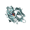
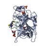
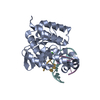
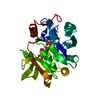
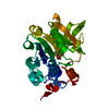
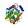
| Entry | Database: PDB / ID: 1jzt | ||||||
|---|---|---|---|---|---|---|---|
| Title | Crystal structure of yeast ynu0, YNL200c | ||||||
 Components Components | Hypothetical 27.5 kDa protein in SPX19-GCR2 intergenic region | ||||||
 Keywords Keywords | structural genomics / unknown function / yeast hypothetical protein / selenomethionine / PSI / Protein Structure Initiative / New York SGX Research Center for Structural Genomics / NYSGXRC | ||||||
| Function / homology |  Function and homology information Function and homology informationNicotinate metabolism / NAD(P)H-hydrate epimerase / NAD(P)HX epimerase activity / nicotinamide nucleotide metabolic process / nucleotide binding / mitochondrion / metal ion binding / cytoplasm Similarity search - Function | ||||||
| Biological species |  | ||||||
| Method |  X-RAY DIFFRACTION / SYNCHROTRON / MAD / Resolution: 1.94 Å X-RAY DIFFRACTION / SYNCHROTRON / MAD / Resolution: 1.94 Å | ||||||
 Authors Authors | Jiang, J.-S. / Manning, N.O. / Burley, S.K. / New York SGX Research Center for Structural Genomics (NYSGXRC) | ||||||
 Citation Citation | Journal: To be Published Title: Crystal Structure of Yeast Hypothetical Protein YNU0_YEAST Authors: Jiang, J.-S. / Manning, N.O. / Eswaramoorthy, S. / Gerchman, S.E. / Kumaran, D. / Kycia, J.H. / Lewis, H.A. / Swaminathan, S. / Studier, F.W. | ||||||
| History |
|
- Structure visualization
| Structure viewer | Molecule: MolmilJmol/JSmol |
|---|
- Downloads & links
-Download
| PDBx/mmCIF format | 1jzt.cif.gz | 114.5 KB | Display | PDBx/mmCIF format |
|---|---|---|---|---|
| PDB format | pdb1jzt.ent.gz | 88.1 KB | Display | PDB format |
| PDBx/mmJSON format | 1jzt.json.gz | Tree view | PDBx/mmJSON format | |
| Others |  Other downloads Other downloads |
-Validation report
| Arichive directory | https://data.pdbj.org/pub/pdb/validation_reports/jz/1jztftp://data.pdbj.org/pub/pdb/validation_reports/jz/1jzt | HTTPS FTP |
|---|
-Related structure data
| Similar structure data | |
|---|---|
| Other databases |


-Links
 PDBj
PDBj- Assembly
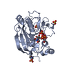
| Deposited unit | 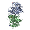
| ||||||||
|---|---|---|---|---|---|---|---|---|---|
| 1 | 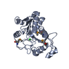
| ||||||||
| 2 | 
| ||||||||
| 3 |
| ||||||||
| Unit cell |
| ||||||||
| Noncrystallographic symmetry (NCS) | NCS oper: (Code: given Matrix: (-0.017708, 0.578797, 0.815279), Vector: |
-Components
| #1: Protein | Mass: 27741.467 Da / Num. of mol.: 2 Source method: isolated from a genetically manipulated source Source: (gene. exp.) Gene: YNL200C / Plasmid: pET 13A / Production host:  #2: Chemical |   Mass: 35.453 Da / Num. of mol.: 2 / Source method: obtained synthetically / Formula: Cl Mass: 35.453 Da / Num. of mol.: 2 / Source method: obtained synthetically / Formula: Cl#3: Water | ChemComp-HOH / |  Mass: 18.015 Da / Num. of mol.: 309 / Source method: isolated from a natural source / Formula: H2O Mass: 18.015 Da / Num. of mol.: 309 / Source method: isolated from a natural source / Formula: H2OHas protein modification | Y | |
|---|
-Experimental details
-Experiment
| Experiment | Method: X-RAY DIFFRACTION / Number of used crystals: 1 |
|---|
- Sample preparation
| Crystal | Density Matthews: 2.29 Å3/Da / Density % sol: 45 % |
|---|---|
| Crystal grow | Temperature: 277 K / Method: vapor diffusion, hanging drop / pH: 8.5 Details: ammonium formate, PEG 3350, pH 8.5, VAPOR DIFFUSION, HANGING DROP, temperature 277K |
-Data collection
| Diffraction | Mean temperature: 100 K | ||||||||||||
|---|---|---|---|---|---|---|---|---|---|---|---|---|---|
| Diffraction source | Source: SYNCHROTRON / Site: NSLS  / Beamline: X12C / Wavelength: 0.9788, 0.9785, 0.9400 / Beamline: X12C / Wavelength: 0.9788, 0.9785, 0.9400 | ||||||||||||
| Detector | Type: BRANDEIS - B1 / Detector: CCD / Date: May 16, 2001 / Details: toroidal mirror | ||||||||||||
| Radiation | Monochromator: Si (111) / Protocol: MAD / Monochromatic (M) / Laue (L): M / Scattering type: x-ray | ||||||||||||
| Radiation wavelength |
| ||||||||||||
| Reflection | Resolution: 1.9→50 Å / Num. all: 40088 / Num. obs: 39366 / % possible obs: 98.2 % / Observed criterion σ(F): 0 / Observed criterion σ(I): 0 / Redundancy: 11 % / Biso Wilson estimate: 8.1 Å2 / Rmerge(I) obs: 0.062 / Net I/σ(I): 12.6 | ||||||||||||
| Reflection shell | Resolution: 1.9→1.97 Å / Redundancy: 9.3 % / Rmerge(I) obs: 0.417 / Mean I/σ(I) obs: 5.8 / % possible all: 92.3 |
- Processing
| Software |
| ||||||||||||||||||||||||||||||||||||
|---|---|---|---|---|---|---|---|---|---|---|---|---|---|---|---|---|---|---|---|---|---|---|---|---|---|---|---|---|---|---|---|---|---|---|---|---|---|
| Refinement | Method to determine structure: MAD / Resolution: 1.94→46.26 Å / Rfactor Rfree error: 0.006 / Data cutoff high absF: 165195.47 / Data cutoff low absF: 0 / Isotropic thermal model: RESTRAINED / Cross valid method: THROUGHOUT / σ(F): 1.5 / Stereochemistry target values: Engh & Huber Details: LOOPS HAVE BEEN MODELLED IN AREAS OF POOR OR MISSING ELECTRON DENSITY. OCCUPANCY IS SET FOR LESS THAN 1 IN AREAS WHERE ELECTRON DENSITY IS AMBIGUOUS, AND IT IS SET EQUAL TO 0 WHERE ELECTRON ...Details: LOOPS HAVE BEEN MODELLED IN AREAS OF POOR OR MISSING ELECTRON DENSITY. OCCUPANCY IS SET FOR LESS THAN 1 IN AREAS WHERE ELECTRON DENSITY IS AMBIGUOUS, AND IT IS SET EQUAL TO 0 WHERE ELECTRON DENSITY IS NONEXISTENT. NOTE PARTICULARLY LOOPS A 119-A 123 AND B 120-B 123. THE FIRST THREE RESIDUES IN EACH CHAIN WERE NOT SEEN IN THE ELECTRON DENSITY.
| ||||||||||||||||||||||||||||||||||||
| Solvent computation | Solvent model: FLAT MODEL / Bsol: 76.6171 Å2 / ksol: 0.392074 e/Å3 | ||||||||||||||||||||||||||||||||||||
| Displacement parameters | Biso mean: 19.1 Å2
| ||||||||||||||||||||||||||||||||||||
| Refine analyze |
| ||||||||||||||||||||||||||||||||||||
| Refinement step | Cycle: LAST / Resolution: 1.94→46.26 Å
| ||||||||||||||||||||||||||||||||||||
| Refine LS restraints |
| ||||||||||||||||||||||||||||||||||||
| Refine LS restraints NCS | NCS model details: CONSTR | ||||||||||||||||||||||||||||||||||||
| LS refinement shell | Resolution: 1.94→2.06 Å / Rfactor Rfree error: 0.015 / Total num. of bins used: 6
| ||||||||||||||||||||||||||||||||||||
| Xplor file |
|
