 Movie
Movie Controller

[English] 日本語
 Yorodumi
- PDB-3qff: Crystal Structure of ADP complex of purK: N5-carboxyaminoimidazol... -
+ Open data

- Basic information
| Entry | Database: PDB / ID: 3qff | ||||||
|---|---|---|---|---|---|---|---|
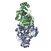
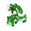
| Title | Crystal Structure of ADP complex of purK: N5-carboxyaminoimidazole ribonucleotide synthetase | ||||||
 Components Components | N5-carboxyaminoimidazole ribonucleotide synthetase | ||||||
 Keywords Keywords | LYASE / carboxylase / ATP binding / ADP binding | ||||||
| Function / homology |  Function and homology information Function and homology informationRossmann fold - #20 / ATP-grasp fold, A domain / ATP-grasp fold, B domain / D-amino Acid Aminotransferase; Chain A, domain 1 / Dna Ligase; domain 1 / Rossmann fold / 2-Layer Sandwich / 3-Layer(aba) Sandwich / Alpha Beta Similarity search - Domain/homology | ||||||
| Biological species |  | ||||||
| Method |  X-RAY DIFFRACTION / SYNCHROTRON / MOLECULAR REPLACEMENT / Resolution: 1.96 Å X-RAY DIFFRACTION / SYNCHROTRON / MOLECULAR REPLACEMENT / Resolution: 1.96 Å | ||||||
 Authors Authors | Fung, L.W. / Tuntland, M.L. / Santarsiero, B.D. / Johnson, M.E. | ||||||
 Citation Citation | Journal: Acta Crystallogr.,Sect.D / Year: 2014 Title: Elucidation of the bicarbonate binding site and insights into the carboxylation mechanism of (N(5))-carboxyaminoimidazole ribonucleotide synthase (PurK) from Bacillus anthracis. Authors: Tuntland, M.L. / Santarsiero, B.D. / Johnson, M.E. / Fung, L.W. | ||||||
| History |
|
- Structure visualization
| Structure viewer | Molecule: MolmilJmol/JSmol |
|---|
- Downloads & links
-Download
| PDBx/mmCIF format | 3qff.cif.gz | 165.6 KB | Display | PDBx/mmCIF format |
|---|---|---|---|---|
| PDB format | pdb3qff.ent.gz | 128.6 KB | Display | PDB format |
| PDBx/mmJSON format | 3qff.json.gz | Tree view | PDBx/mmJSON format | |
| Others |  Other downloads Other downloads |
-Validation report
| Arichive directory | https://data.pdbj.org/pub/pdb/validation_reports/qf/3qffftp://data.pdbj.org/pub/pdb/validation_reports/qf/3qff | HTTPS FTP |
|---|
-Related structure data
| Related structure data |  3r5hC 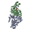 3v4sC 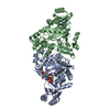 4dlkC  3q2oS S: Starting model for refinement C: citing same article ( |
|---|---|
| Similar structure data |


-Links
 PDBj
PDBj- Assembly
| Deposited unit | 
| ||||||||
|---|---|---|---|---|---|---|---|---|---|
| 1 |
| ||||||||
| Unit cell |
|
-Components
| #1: Protein | Mass: 43209.355 Da / Num. of mol.: 2 Source method: isolated from a genetically manipulated source Source: (gene. exp.) References: UniProt: C3PBM5, phosphoribosylaminoimidazole carboxylase #2: Chemical |   Mass: 427.201 Da / Num. of mol.: 2 / Source method: obtained synthetically / Formula: C10H15N5O10P2 / Comment: ADP, energy-carrying molecule*YM Mass: 427.201 Da / Num. of mol.: 2 / Source method: obtained synthetically / Formula: C10H15N5O10P2 / Comment: ADP, energy-carrying molecule*YM#3: Water | ChemComp-HOH / |  Mass: 18.015 Da / Num. of mol.: 317 / Source method: isolated from a natural source / Formula: H2O Mass: 18.015 Da / Num. of mol.: 317 / Source method: isolated from a natural source / Formula: H2O |
|---|
-Experimental details
-Experiment
| Experiment | Method: X-RAY DIFFRACTION / Number of used crystals: 1 |
|---|
- Sample preparation
| Crystal | Density Matthews: 2.27 Å3/Da / Density % sol: 45.91 % |
|---|---|
| Crystal grow | Temperature: 298 K / Method: vapor diffusion, hanging drop / pH: 6.5 Details: 0.1M MES, 5mM DTT, 9% glycerol, 10% PEG6000, Hampton Research Silver Bullet Bio additive C3/27 and 3mM ADP, pH 6.5, vapor diffusion, hanging drop, temperature 298K |
-Data collection
| Diffraction | Mean temperature: 100 K | ||||||||||||||||||||||||||||||||||||||||||||||||||||||||||||||||||||||
|---|---|---|---|---|---|---|---|---|---|---|---|---|---|---|---|---|---|---|---|---|---|---|---|---|---|---|---|---|---|---|---|---|---|---|---|---|---|---|---|---|---|---|---|---|---|---|---|---|---|---|---|---|---|---|---|---|---|---|---|---|---|---|---|---|---|---|---|---|---|---|---|
| Diffraction source | Source: SYNCHROTRON / Site: APS  / Beamline: 22-ID / Wavelength: 1 Å / Beamline: 22-ID / Wavelength: 1 Å | ||||||||||||||||||||||||||||||||||||||||||||||||||||||||||||||||||||||
| Detector | Type: MARMOSAIC 300 mm CCD / Detector: CCD / Date: Dec 2, 2010 | ||||||||||||||||||||||||||||||||||||||||||||||||||||||||||||||||||||||
| Radiation | Protocol: SINGLE WAVELENGTH / Monochromatic (M) / Laue (L): M / Scattering type: x-ray | ||||||||||||||||||||||||||||||||||||||||||||||||||||||||||||||||||||||
| Radiation wavelength | Wavelength: 1 Å / Relative weight: 1 | ||||||||||||||||||||||||||||||||||||||||||||||||||||||||||||||||||||||
| Reflection | Highest resolution: 1.96 Å / Num. obs: 55699 / % possible obs: 97.1 % / Observed criterion σ(I): -3 / Biso Wilson estimate: 39.42 Å2 / Rmerge(I) obs: 0.091 / Net I/σ(I): 14.43 | ||||||||||||||||||||||||||||||||||||||||||||||||||||||||||||||||||||||
| Reflection shell | Diffraction-ID: 1
|
- Processing
| Software |
| |||||||||||||||||||||||||||||||||||||||||||||||||||||||||||||||||
|---|---|---|---|---|---|---|---|---|---|---|---|---|---|---|---|---|---|---|---|---|---|---|---|---|---|---|---|---|---|---|---|---|---|---|---|---|---|---|---|---|---|---|---|---|---|---|---|---|---|---|---|---|---|---|---|---|---|---|---|---|---|---|---|---|---|---|
| Refinement | Method to determine structure: MOLECULAR REPLACEMENT Starting model: PDB entry 3Q2O Resolution: 1.96→19.97 Å / Cor.coef. Fo:Fc: 0.954 / Cor.coef. Fo:Fc free: 0.926 / WRfactor Rfree: 0.2551 / WRfactor Rwork: 0.1987 / Occupancy max: 1 / Occupancy min: 0.3 / FOM work R set: 0.841 / SU B: 4.24 / SU ML: 0.121 / SU R Cruickshank DPI: 0.1901 / SU Rfree: 0.1744 / Cross valid method: THROUGHOUT / σ(F): 0 / ESU R Free: 0.174 / Stereochemistry target values: MAXIMUM LIKELIHOOD Details: HYDROGENS HAVE BEEN ADDED IN THE RIDING POSITIONS U VALUES : REFINED INDIVIDUALLY
| |||||||||||||||||||||||||||||||||||||||||||||||||||||||||||||||||
| Solvent computation | Ion probe radii: 0.8 Å / Shrinkage radii: 0.8 Å / VDW probe radii: 1.4 Å / Solvent model: MASK | |||||||||||||||||||||||||||||||||||||||||||||||||||||||||||||||||
| Displacement parameters | Biso max: 68.43 Å2 / Biso mean: 34.6697 Å2 / Biso min: 12.81 Å2
| |||||||||||||||||||||||||||||||||||||||||||||||||||||||||||||||||
| Refinement step | Cycle: LAST / Resolution: 1.96→19.97 Å
| |||||||||||||||||||||||||||||||||||||||||||||||||||||||||||||||||
| Refine LS restraints |
| |||||||||||||||||||||||||||||||||||||||||||||||||||||||||||||||||
| LS refinement shell | Resolution: 1.961→2.012 Å / Total num. of bins used: 20
|
