 Movie
Movie Controller


+ Open data

- Basic information
| Entry | Database: PDB / ID: 3q8e | ||||||
|---|---|---|---|---|---|---|---|
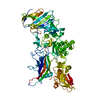
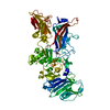
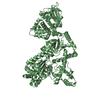
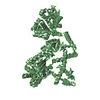
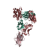
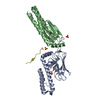
| Title | Crystal structure of Protective Antigen W346F (pH 8.5) | ||||||
 Components Components | Protective antigen | ||||||
 Keywords Keywords | TOXIN / Protective Antigen / Anthrax / pH stability | ||||||
| Function / homology |  Function and homology information Function and homology informationsymbiont-mediated suppression of host MAPK cascade / host cell cytosol / Uptake and function of anthrax toxins / host cell endosome membrane / protein homooligomerization / toxin activity / host cell plasma membrane / extracellular region / metal ion binding / identical protein binding Similarity search - Function | ||||||
| Biological species |  | ||||||
| Method |  X-RAY DIFFRACTION / SYNCHROTRON / FOURIER SYNTHESIS / Resolution: 2.098 Å X-RAY DIFFRACTION / SYNCHROTRON / FOURIER SYNTHESIS / Resolution: 2.098 Å | ||||||
 Authors Authors | Lovell, S. / Battaile, K.P. / Rajapaksha, M. / Janowiak, B.E. / Andra, K.K. / Bann, J.G. | ||||||
 Citation Citation | Journal: Protein Sci. / Year: 2012 Title: pH effects on binding between the anthrax protective antigen and the host cellular receptor CMG2. Authors: Rajapaksha, M. / Lovell, S. / Janowiak, B.E. / Andra, K.K. / Battaile, K.P. / Bann, J.G. | ||||||
| History |
|
- Structure visualization
| Structure viewer | Molecule: MolmilJmol/JSmol |
|---|
- Downloads & links
-Download
| PDBx/mmCIF format | 3q8e.cif.gz | 154.2 KB | Display | PDBx/mmCIF format |
|---|---|---|---|---|
| PDB format | pdb3q8e.ent.gz | 118.1 KB | Display | PDB format |
| PDBx/mmJSON format | 3q8e.json.gz | Tree view | PDBx/mmJSON format | |
| Others |  Other downloads Other downloads |
-Validation report
| Arichive directory | https://data.pdbj.org/pub/pdb/validation_reports/q8/3q8eftp://data.pdbj.org/pub/pdb/validation_reports/q8/3q8e | HTTPS FTP |
|---|
-Related structure data
| Related structure data |  3q8aC  3q8bC  3q8cC  3q8fC  3mhzS C: citing same article ( S: Starting model for refinement |
|---|---|
| Similar structure data |


-Links
 PDBj
PDBj


- Assembly
| Deposited unit | 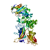
| ||||||||
|---|---|---|---|---|---|---|---|---|---|
| 1 |
| ||||||||
| Unit cell |
|
-Components
| #1: Protein | Mass: 82729.789 Da / Num. of mol.: 1 / Mutation: W346F Source method: isolated from a genetically manipulated source Source: (gene. exp.) | ||
|---|---|---|---|
| #2: Chemical |   Mass: 40.078 Da / Num. of mol.: 2 / Source method: obtained synthetically / Formula: Ca Mass: 40.078 Da / Num. of mol.: 2 / Source method: obtained synthetically / Formula: Ca#3: Water | ChemComp-HOH / |  Mass: 18.015 Da / Num. of mol.: 243 / Source method: isolated from a natural source / Formula: H2O Mass: 18.015 Da / Num. of mol.: 243 / Source method: isolated from a natural source / Formula: H2O |
-Experimental details
-Experiment
| Experiment | Method: X-RAY DIFFRACTION / Number of used crystals: 1 |
|---|
- Sample preparation
| Crystal | Density Matthews: 2.37 Å3/Da / Density % sol: 48.02 % |
|---|---|
| Crystal grow | Temperature: 293 K / Method: vapor diffusion / pH: 8.5 Details: 25%(w/v) PEG 1500, 100mM SPG Buffer, pH 8.5, vapor diffusion, temperature 293K |
-Data collection
| Diffraction | Mean temperature: 100 K | ||||||||||||||||||||||||||||||||||||||||||||||||||||||||||||||||||||||||||||||||||||||||
|---|---|---|---|---|---|---|---|---|---|---|---|---|---|---|---|---|---|---|---|---|---|---|---|---|---|---|---|---|---|---|---|---|---|---|---|---|---|---|---|---|---|---|---|---|---|---|---|---|---|---|---|---|---|---|---|---|---|---|---|---|---|---|---|---|---|---|---|---|---|---|---|---|---|---|---|---|---|---|---|---|---|---|---|---|---|---|---|---|---|
| Diffraction source | Source: SYNCHROTRON / Site: APS  / Beamline: 17-ID / Wavelength: 1 Å / Beamline: 17-ID / Wavelength: 1 Å | ||||||||||||||||||||||||||||||||||||||||||||||||||||||||||||||||||||||||||||||||||||||||
| Detector | Type: DECTRIS PILATUS 6M / Detector: PIXEL / Date: Jan 1, 2010 | ||||||||||||||||||||||||||||||||||||||||||||||||||||||||||||||||||||||||||||||||||||||||
| Radiation | Protocol: SINGLE WAVELENGTH / Scattering type: x-ray | ||||||||||||||||||||||||||||||||||||||||||||||||||||||||||||||||||||||||||||||||||||||||
| Radiation wavelength | Wavelength: 1 Å / Relative weight: 1 | ||||||||||||||||||||||||||||||||||||||||||||||||||||||||||||||||||||||||||||||||||||||||
| Reflection | Resolution: 2.098→117.205 Å / Num. all: 46703 / Num. obs: 46703 / % possible obs: 100 % / Observed criterion σ(F): 0 / Observed criterion σ(I): 0 / Redundancy: 6.3 % / Rsym value: 0.06 / Net I/σ(I): 17.7 | ||||||||||||||||||||||||||||||||||||||||||||||||||||||||||||||||||||||||||||||||||||||||
| Reflection shell | Diffraction-ID: 1
|
- Processing
| Software |
| |||||||||||||||||||||||||||||||||||||||||||||||||||||||||||||||||||||||||||||
|---|---|---|---|---|---|---|---|---|---|---|---|---|---|---|---|---|---|---|---|---|---|---|---|---|---|---|---|---|---|---|---|---|---|---|---|---|---|---|---|---|---|---|---|---|---|---|---|---|---|---|---|---|---|---|---|---|---|---|---|---|---|---|---|---|---|---|---|---|---|---|---|---|---|---|---|---|---|---|
| Refinement | Method to determine structure: FOURIER SYNTHESIS Starting model: PDB entry 3MHZ Resolution: 2.098→45.291 Å / Occupancy max: 1 / Occupancy min: 1 / SU ML: 0.31 / σ(F): 1.35 / Phase error: 27.55 / Stereochemistry target values: ML
| |||||||||||||||||||||||||||||||||||||||||||||||||||||||||||||||||||||||||||||
| Solvent computation | Shrinkage radii: 0.77 Å / VDW probe radii: 0.9 Å / Solvent model: FLAT BULK SOLVENT MODEL / Bsol: 42.297 Å2 / ksol: 0.383 e/Å3 | |||||||||||||||||||||||||||||||||||||||||||||||||||||||||||||||||||||||||||||
| Displacement parameters | Biso max: 118.48 Å2 / Biso mean: 44.9292 Å2 / Biso min: 20.23 Å2
| |||||||||||||||||||||||||||||||||||||||||||||||||||||||||||||||||||||||||||||
| Refinement step | Cycle: LAST / Resolution: 2.098→45.291 Å
| |||||||||||||||||||||||||||||||||||||||||||||||||||||||||||||||||||||||||||||
| Refine LS restraints |
| |||||||||||||||||||||||||||||||||||||||||||||||||||||||||||||||||||||||||||||
| LS refinement shell | Refine-ID: X-RAY DIFFRACTION / Total num. of bins used: 10
|
