 Movie
Movie Controller

[English] 日本語
 Yorodumi
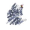
- PDB-3q79: Cryptococcus neoformans protein farnesyltransferase in complex wi... -
+ Open data

- Basic information
| Entry | Database: PDB / ID: 3q79 | ||||||
|---|---|---|---|---|---|---|---|
| Title | Cryptococcus neoformans protein farnesyltransferase in complex with farnesyl-DDPTASACNIQ product | ||||||
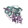
 Components Components |
| ||||||
 Keywords Keywords | TRANSFERASE / protein prenyltransferase | ||||||
| Function / homology |  Function and homology information Function and homology informationprotein prenyltransferase activity / protein farnesyltransferase / protein farnesyltransferase activity / protein farnesyltransferase complex / zinc ion binding Similarity search - Function | ||||||
| Biological species |  Cryptococcus neoformans (Cryptococcus neoformans serotype A) Cryptococcus neoformans (Cryptococcus neoformans serotype A) | ||||||
| Method |  X-RAY DIFFRACTION / SYNCHROTRON / MOLECULAR REPLACEMENT / Resolution: 2.506 Å X-RAY DIFFRACTION / SYNCHROTRON / MOLECULAR REPLACEMENT / Resolution: 2.506 Å | ||||||
 Authors Authors | Hast, M.A. / Beese, L.S. | ||||||
 Citation Citation | Journal: J.Biol.Chem. / Year: 2011 Title: Structures of Cryptococcus neoformans Protein Farnesyltransferase Reveal Strategies for Developing Inhibitors That Target Fungal Pathogens. Authors: Hast, M.A. / Nichols, C.B. / Armstrong, S.M. / Kelly, S.M. / Hellinga, H.W. / Alspaugh, J.A. / Beese, L.S. | ||||||
| History |
| ||||||
| Remark 999 | There is no UniPort database reference sequence available at the time of the deposition. The ...There is no UniPort database reference sequence available at the time of the deposition. The sequences presented here are of farnesyltransferase alpha subunit(residues 15-349) and beta subunit from Cryptococcus neoformans var. grubii strain H99. The first 14 residues in the alpha subunit(chain A) represent expression tag. |
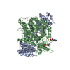
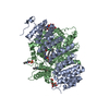
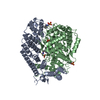
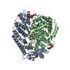
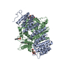
- Structure visualization
| Structure viewer | Molecule: MolmilJmol/JSmol |
|---|
- Downloads & links
-Download
| PDBx/mmCIF format | 3q79.cif.gz | 339 KB | Display | PDBx/mmCIF format |
|---|---|---|---|---|
| PDB format | pdb3q79.ent.gz | 273.2 KB | Display | PDB format |
| PDBx/mmJSON format | 3q79.json.gz | Tree view | PDBx/mmJSON format | |
| Others |  Other downloads Other downloads |
-Validation report
| Arichive directory | https://data.pdbj.org/pub/pdb/validation_reports/q7/3q79ftp://data.pdbj.org/pub/pdb/validation_reports/q7/3q79 | HTTPS FTP |
|---|
-Related structure data
| Related structure data |  3q73C 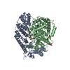 3q75C  3q78C  3q7aC  3q7fC  3sfxC 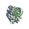 3sfyC C: citing same article ( |
|---|---|
| Similar structure data |


-Links
 PDBj
PDBj

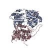
- Assembly
| Deposited unit | 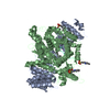
| ||||||||
|---|---|---|---|---|---|---|---|---|---|
| 1 |
| ||||||||
| Unit cell |
| ||||||||
| Components on special symmetry positions |
|
-Components
-Farnesyltransferase ... , 2 types, 2 molecules AB
| #1: Protein | Mass: 40913.234 Da / Num. of mol.: 1 Source method: isolated from a genetically manipulated source Source: (gene. exp.) Cryptococcus neoformans (Cryptococcus neoformans serotype A)Strain: H99 / Production host:  |
|---|---|
| #2: Protein | Mass: 56806.879 Da / Num. of mol.: 1 Source method: isolated from a genetically manipulated source Source: (gene. exp.) Cryptococcus neoformans (Cryptococcus neoformans serotype A)Strain: H99 / Production host: |
-Protein/peptide , 1 types, 1 molecules P
| #3: Protein/peptide | Mass: 1134.175 Da / Num. of mol.: 1 / Source method: obtained synthetically / Details: synthetic substrate |
|---|
-Non-polymers , 5 types, 389 molecules 


| #4: Chemical | ChemComp-ZN /  Mass: 65.409 Da / Num. of mol.: 1 / Source method: obtained synthetically / Formula: Zn Mass: 65.409 Da / Num. of mol.: 1 / Source method: obtained synthetically / Formula: Zn | ||
|---|---|---|---|
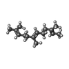
| #5: Chemical | ChemComp-FAR /  Mass: 206.367 Da / Num. of mol.: 1 / Source method: obtained synthetically / Formula: C15H26 Mass: 206.367 Da / Num. of mol.: 1 / Source method: obtained synthetically / Formula: C15H26 | ||
| #6: Chemical | ChemComp-SO4 /  Mass: 96.063 Da / Num. of mol.: 1 / Source method: obtained synthetically / Formula: SO4 Mass: 96.063 Da / Num. of mol.: 1 / Source method: obtained synthetically / Formula: SO4 | ||
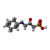
| #7: Chemical |  Mass: 237.316 Da / Num. of mol.: 3 / Source method: obtained synthetically / Formula: C9H19NO4S Mass: 237.316 Da / Num. of mol.: 3 / Source method: obtained synthetically / Formula: C9H19NO4S#8: Water | ChemComp-HOH / | Mass: 18.015 Da / Num. of mol.: 383 / Source method: isolated from a natural source / Formula: H2O |
-Details
| Has protein modification | Y |
|---|
-Experimental details
-Experiment
| Experiment | Method: X-RAY DIFFRACTION / Number of used crystals: 1 |
|---|
- Sample preparation
| Crystal | Density Matthews: 3.31 Å3/Da / Density % sol: 62.88 % |
|---|---|
| Crystal grow | Temperature: 290 K / Method: vapor diffusion, hanging drop / pH: 9.5 Details: 15% PEG 4K, 100mM CAPSO pH 9.5, 100mM NaCl, 150mM Lithium sulfate, VAPOR DIFFUSION, HANGING DROP, temperature 290K |
-Data collection
| Diffraction | Mean temperature: 100 K |
|---|---|
| Diffraction source | Source: SYNCHROTRON / Site: ALS  / Beamline: 12.3.1 / Wavelength: 0.9795 Å / Beamline: 12.3.1 / Wavelength: 0.9795 Å |
| Detector | Type: ADSC QUANTUM 315 / Detector: CCD / Date: Apr 21, 2010 |
| Radiation | Protocol: SINGLE WAVELENGTH / Monochromatic (M) / Laue (L): M / Scattering type: x-ray |
| Radiation wavelength | Wavelength: 0.9795 Å / Relative weight: 1 |
| Reflection | Resolution: 2.5→50 Å / Num. obs: 44750 / % possible obs: 96.3 % / Observed criterion σ(I): -3 |
| Reflection shell | Resolution: 2.5→2.6 Å / % possible all: 66.8 |
- Processing
| Software |
| |||||||||||||||||||||||||||||||||||||||||||||||||||||||||||||||||||||||||||||
|---|---|---|---|---|---|---|---|---|---|---|---|---|---|---|---|---|---|---|---|---|---|---|---|---|---|---|---|---|---|---|---|---|---|---|---|---|---|---|---|---|---|---|---|---|---|---|---|---|---|---|---|---|---|---|---|---|---|---|---|---|---|---|---|---|---|---|---|---|---|---|---|---|---|---|---|---|---|---|
| Refinement | Method to determine structure: MOLECULAR REPLACEMENT / Resolution: 2.506→47.954 Å / SU ML: 0.35 / σ(F): 1.99 / Phase error: 26.37 / Stereochemistry target values: ML
| |||||||||||||||||||||||||||||||||||||||||||||||||||||||||||||||||||||||||||||
| Solvent computation | Shrinkage radii: 0.9 Å / VDW probe radii: 1.11 Å / Solvent model: FLAT BULK SOLVENT MODEL / Bsol: 40.64 Å2 / ksol: 0.335 e/Å3 | |||||||||||||||||||||||||||||||||||||||||||||||||||||||||||||||||||||||||||||
| Displacement parameters |
| |||||||||||||||||||||||||||||||||||||||||||||||||||||||||||||||||||||||||||||
| Refinement step | Cycle: LAST / Resolution: 2.506→47.954 Å
| |||||||||||||||||||||||||||||||||||||||||||||||||||||||||||||||||||||||||||||
| Refine LS restraints |
| |||||||||||||||||||||||||||||||||||||||||||||||||||||||||||||||||||||||||||||
| LS refinement shell |
| |||||||||||||||||||||||||||||||||||||||||||||||||||||||||||||||||||||||||||||
| Refinement TLS params. | Method: refined / Origin x: 28.3434 Å / Origin y: -31.4456 Å / Origin z: -2.6667 Å
| |||||||||||||||||||||||||||||||||||||||||||||||||||||||||||||||||||||||||||||
| Refinement TLS group | Selection details: all |
