 Movie
Movie Controller

[English] 日本語
 Yorodumi
- PDB-3q4a: Crystal structure of the TPR domain of CHIP complexed with phosph... -
+ Open data

- Basic information
| Entry | Database: PDB / ID: 3q4a | ||||||
|---|---|---|---|---|---|---|---|
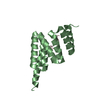
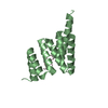
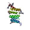
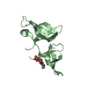
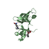
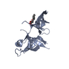
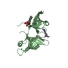
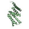
| Title | Crystal structure of the TPR domain of CHIP complexed with phosphorylated Smad1 peptide | ||||||
 Components Components |
| ||||||
 Keywords Keywords | LIGASE/TRANSCRIPTION / E3 ubiquitin ligase / LIGASE-TRANSCRIPTION complex | ||||||
| Function / homology |  Function and homology information Function and homology informationmesodermal cell fate commitment / homomeric SMAD protein complex / osteoblast fate commitment / anti-Mullerian hormone receptor signaling pathway / positive regulation of chaperone-mediated protein complex assembly / SMAD protein complex / Downregulation of TGF-beta receptor signaling / RUNX2 regulates bone development / regulation of glucocorticoid metabolic process / heteromeric SMAD protein complex ...mesodermal cell fate commitment / homomeric SMAD protein complex / osteoblast fate commitment / anti-Mullerian hormone receptor signaling pathway / positive regulation of chaperone-mediated protein complex assembly / SMAD protein complex / Downregulation of TGF-beta receptor signaling / RUNX2 regulates bone development / regulation of glucocorticoid metabolic process / heteromeric SMAD protein complex / co-SMAD binding / negative regulation of vascular associated smooth muscle contraction / Downregulation of ERBB2 signaling / negative regulation of muscle cell differentiation / negative regulation of peroxisome proliferator activated receptor signaling pathway / Regulation of PTEN stability and activity / positive regulation of cartilage development / Regulation of TNFR1 signaling / Regulation of necroptotic cell death / DEAD/H-box RNA helicase binding / Regulation of RUNX2 expression and activity / ubiquitin conjugating enzyme complex / positive regulation of ERAD pathway / primary miRNA binding / gamete generation / positive regulation of smooth muscle cell apoptotic process / hindbrain development / positive regulation of mitophagy / negative regulation of cardiac muscle hypertrophy / cardiac conduction system development / primary miRNA processing / nuclear inclusion body / Signaling by BMP / SMAD protein signal transduction / embryonic pattern specification / misfolded protein binding / I-SMAD binding / cartilage development / Antigen processing: Ubiquitination & Proteasome degradation / Cardiogenesis / cellular response to misfolded protein / positive regulation of dendrite development / nuclear inner membrane / ureteric bud development / ubiquitin-ubiquitin ligase activity / chaperone-mediated autophagy / positive regulation of sprouting angiogenesis / midbrain development / TPR domain binding / SMAD binding / positive regulation of proteolysis / negative regulation of smooth muscle cell apoptotic process / R-SMAD binding / protein quality control for misfolded or incompletely synthesized proteins / protein folding chaperone complex / anatomical structure morphogenesis / positive regulation of osteoblast differentiation / cardiac muscle cell proliferation / BMP signaling pathway / protein K63-linked ubiquitination / protein monoubiquitination / ubiquitin ligase complex / Transcriptional regulation of brown and beige adipocyte differentiation by EBF2 / protein autoubiquitination / endoplasmic reticulum unfolded protein response / heat shock protein binding / ERAD pathway / transforming growth factor beta receptor signaling pathway / Hsp70 protein binding / ossification / response to ischemia / positive regulation of protein ubiquitination / stem cell differentiation / negative regulation of transforming growth factor beta receptor signaling pathway / regulation of protein stability / Hsp90 protein binding / RING-type E3 ubiquitin transferase / bone development / positive regulation of miRNA transcription / G protein-coupled receptor binding / Z disc / kinase binding / protein polyubiquitination / ubiquitin-protein transferase activity / osteoblast differentiation / ubiquitin protein ligase activity / MAPK cascade / positive regulation of proteasomal ubiquitin-dependent protein catabolic process / cellular response to heat / protein folding / protein-folding chaperone binding / DNA-binding transcription activator activity, RNA polymerase II-specific / transcription by RNA polymerase II / cellular response to hypoxia / ubiquitin-dependent protein catabolic process / intracellular iron ion homeostasis / proteasome-mediated ubiquitin-dependent protein catabolic process / protein-macromolecule adaptor activity / DNA-binding transcription factor activity, RNA polymerase II-specific / cell differentiation Similarity search - Function | ||||||
| Biological species |   Homo sapiens (human) Homo sapiens (human) | ||||||
| Method |  X-RAY DIFFRACTION / SYNCHROTRON / MOLECULAR REPLACEMENT / Resolution: 1.542 Å X-RAY DIFFRACTION / SYNCHROTRON / MOLECULAR REPLACEMENT / Resolution: 1.542 Å | ||||||
 Authors Authors | Wang, L. / Chen, L. / Wu, J.W. | ||||||
 Citation Citation | Journal: J.Biol.Chem. / Year: 2011 Title: Molecular Mechanism of the Negative Regulation of Smad1/5 Protein by Carboxyl Terminus of Hsc70-interacting Protein (CHIP). Authors: Wang, L. / Liu, Y.T. / Hao, R. / Chen, L. / Chang, Z. / Wang, H.R. / Wang, Z.X. / Wu, J.W. | ||||||
| History |
|
- Structure visualization
| Structure viewer | Molecule: MolmilJmol/JSmol |
|---|
- Downloads & links
-Download
| PDBx/mmCIF format | 3q4a.cif.gz | 73.3 KB | Display | PDBx/mmCIF format |
|---|---|---|---|---|
| PDB format | pdb3q4a.ent.gz | 54.1 KB | Display | PDB format |
| PDBx/mmJSON format | 3q4a.json.gz | Tree view | PDBx/mmJSON format | |
| Others |  Other downloads Other downloads |
-Validation report
| Arichive directory | https://data.pdbj.org/pub/pdb/validation_reports/q4/3q4aftp://data.pdbj.org/pub/pdb/validation_reports/q4/3q4a | HTTPS FTP |
|---|
-Related structure data
| Related structure data |  3q47C  3q49C 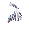 2c2lS C: citing same article ( S: Starting model for refinement |
|---|---|
| Similar structure data |


-Links
 PDBj
PDBj


- Assembly
| Deposited unit | 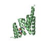
| ||||||||
|---|---|---|---|---|---|---|---|---|---|
| 1 |
| ||||||||
| Unit cell |
|
-Components
| #1: Protein | Mass: 15733.964 Da / Num. of mol.: 1 / Fragment: TPR domain Source method: isolated from a genetically manipulated source Source: (gene. exp.)  References: UniProt: Q9WUD1, Ligases; Forming carbon-nitrogen bonds; Acid-amino-acid ligases (peptide synthases) |
|---|---|
| #2: Protein/peptide | Mass: 1185.052 Da / Num. of mol.: 1 / Source method: obtained synthetically / Details: The peptide was chemically synthesized. / Source: (synth.) Homo sapiens (human) / References: UniProt: Q15797*PLUS |
| #3: Water | ChemComp-HOH /  Mass: 18.015 Da / Num. of mol.: 185 / Source method: isolated from a natural source / Formula: H2O Mass: 18.015 Da / Num. of mol.: 185 / Source method: isolated from a natural source / Formula: H2O |
| Has protein modification | Y |
-Experimental details
-Experiment
| Experiment | Method: X-RAY DIFFRACTION / Number of used crystals: 1 |
|---|
- Sample preparation
| Crystal | Density Matthews: 1.92 Å3/Da / Density % sol: 36.09 % |
|---|---|
| Crystal grow | Temperature: 294 K / Method: vapor diffusion, hanging drop / pH: 7 Details: 100mM Bis-tris propane pH 7.0, 40% PEG MME 2000, 1% PEG MME 550, 50mM magnesium acetate, VAPOR DIFFUSION, HANGING DROP, temperature 294K |
-Data collection
| Diffraction | Mean temperature: 100 K |
|---|---|
| Diffraction source | Source: SYNCHROTRON / Site: SSRF  / Beamline: BL17U / Wavelength: 0.97924 Å / Beamline: BL17U / Wavelength: 0.97924 Å |
| Detector | Type: MARMOSAIC 225 mm CCD / Detector: CCD / Date: Jul 12, 2009 |
| Radiation | Monochromator: GRAPHITE / Protocol: SINGLE WAVELENGTH / Monochromatic (M) / Laue (L): M / Scattering type: x-ray |
| Radiation wavelength | Wavelength: 0.97924 Å / Relative weight: 1 |
| Reflection | Resolution: 1.54→33 Å / Num. all: 19828 / Num. obs: 19797 / % possible obs: 99.4 % / Observed criterion σ(F): 0 / Observed criterion σ(I): 0 / Redundancy: 7.7 % / Rmerge(I) obs: 0.053 / Net I/σ(I): 57.7 |
| Reflection shell | Resolution: 1.54→1.57 Å / Redundancy: 7.1 % / Rmerge(I) obs: 0.171 / Mean I/σ(I) obs: 11.8 / % possible all: 97.9 |
- Processing
| Software |
| ||||||||||||||||||||||||||||||||||||||||||||||||
|---|---|---|---|---|---|---|---|---|---|---|---|---|---|---|---|---|---|---|---|---|---|---|---|---|---|---|---|---|---|---|---|---|---|---|---|---|---|---|---|---|---|---|---|---|---|---|---|---|---|
| Refinement | Method to determine structure: MOLECULAR REPLACEMENT Starting model: 2C2L Resolution: 1.542→32.984 Å / SU ML: 0.16 / σ(F): 1.34 / Stereochemistry target values: ML
| ||||||||||||||||||||||||||||||||||||||||||||||||
| Solvent computation | Shrinkage radii: 0.9 Å / VDW probe radii: 1.11 Å / Solvent model: FLAT BULK SOLVENT MODEL / Bsol: 82.234 Å2 / ksol: 0.379 e/Å3 | ||||||||||||||||||||||||||||||||||||||||||||||||
| Displacement parameters |
| ||||||||||||||||||||||||||||||||||||||||||||||||
| Refinement step | Cycle: LAST / Resolution: 1.542→32.984 Å
| ||||||||||||||||||||||||||||||||||||||||||||||||
| Refine LS restraints |
| ||||||||||||||||||||||||||||||||||||||||||||||||
| LS refinement shell | Refine-ID: X-RAY DIFFRACTION / Total num. of bins used: 7
|
