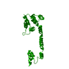
 Movie
Movie Controller

[English] 日本語
 Yorodumi
- PDB-3px2: Structure of TylM1 from Streptomyces fradiae H123N mutant in comp... -
+ Open data

- Basic information
| Entry | Database: PDB / ID: 3px2 | ||||||
|---|---|---|---|---|---|---|---|
| Title | Structure of TylM1 from Streptomyces fradiae H123N mutant in complex with SAH and dTDP-Quip3N | ||||||
 Components Components | N-methyltransferase | ||||||
 Keywords Keywords | TRANSFERASE / SAM binding / N / N-dimethyltransferase / dTDP-Quip3N binding | ||||||
| Function / homology |  Function and homology information Function and homology informationdTDP-3-amino-3,6-dideoxy-alpha-D-glucopyranose N,N-dimethyltransferase / S-adenosylmethionine-dependent methyltransferase activity / antibiotic biosynthetic process / methylation / protein homodimerization activity Similarity search - Function | ||||||
| Biological species |  Streptomyces fradiae (bacteria) Streptomyces fradiae (bacteria) | ||||||
| Method |  X-RAY DIFFRACTION / MOLECULAR REPLACEMENT / Resolution: 1.65 Å X-RAY DIFFRACTION / MOLECULAR REPLACEMENT / Resolution: 1.65 Å | ||||||
 Authors Authors | Holden, H.M. / Carney, A.E. | ||||||
 Citation Citation | Journal: Biochemistry / Year: 2011 Title: Molecular Architecture of TylM1 from Streptomyces fradiae: An N,N-Dimethyltransferase Involved in the Production of dTDP-d-mycaminose . Authors: Carney, A.E. / Holden, H.M. | ||||||
| History |
|
- Structure visualization
| Structure viewer | Molecule: MolmilJmol/JSmol |
|---|
- Downloads & links
-Download
| PDBx/mmCIF format | 3px2.cif.gz | 118.5 KB | Display | PDBx/mmCIF format |
|---|---|---|---|---|
| PDB format | pdb3px2.ent.gz | 90.5 KB | Display | PDB format |
| PDBx/mmJSON format | 3px2.json.gz | Tree view | PDBx/mmJSON format | |
| Others |  Other downloads Other downloads |
-Validation report
| Summary document | 3px2_validation.pdf.gz | 1.6 MB | Display | wwPDB validaton report |
|---|---|---|---|---|
| Full document | 3px2_full_validation.pdf.gz | 1.6 MB | Display | |
| Data in XML | 3px2_validation.xml.gz | 26.1 KB | Display | |
| Data in CIF | 3px2_validation.cif.gz | 37.5 KB | Display | |
| Arichive directory | https://data.pdbj.org/pub/pdb/validation_reports/px/3px2ftp://data.pdbj.org/pub/pdb/validation_reports/px/3px2 | HTTPS FTP |
-Related structure data
| Related structure data |  3pfgC  3pfhSC  3px3C C: citing same article ( S: Starting model for refinement |
|---|---|
| Similar structure data |


-Links
 PDBj
PDBj- Assembly
| Deposited unit | 
| ||||||||
|---|---|---|---|---|---|---|---|---|---|
| 1 |
| ||||||||
| Unit cell |
|
-Components
| #1: Protein | Mass: 28375.697 Da / Num. of mol.: 2 / Mutation: H123N Source method: isolated from a genetically manipulated source Source: (gene. exp.) Streptomyces fradiae (bacteria) / Strain: ATCC 19609 / Gene: tylM1, tylMI(orf3*) / Plasmid: pET31b / Production host: #2: Chemical |   Type: L-peptide linking / Mass: 384.411 Da / Num. of mol.: 2 / Source method: obtained synthetically / Formula: C14H20N6O5S Type: L-peptide linking / Mass: 384.411 Da / Num. of mol.: 2 / Source method: obtained synthetically / Formula: C14H20N6O5S#3: Chemical | 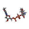  Mass: 547.345 Da / Num. of mol.: 2 / Source method: obtained synthetically / Formula: C16H27N3O14P2 Mass: 547.345 Da / Num. of mol.: 2 / Source method: obtained synthetically / Formula: C16H27N3O14P2#4: Chemical |   Mass: 62.068 Da / Num. of mol.: 2 / Source method: obtained synthetically / Formula: C2H6O2 Mass: 62.068 Da / Num. of mol.: 2 / Source method: obtained synthetically / Formula: C2H6O2#5: Water | ChemComp-HOH / |  Mass: 18.015 Da / Num. of mol.: 414 / Source method: isolated from a natural source / Formula: H2O Mass: 18.015 Da / Num. of mol.: 414 / Source method: isolated from a natural source / Formula: H2O |
|---|
-Experimental details
-Experiment
| Experiment | Method: X-RAY DIFFRACTION / Number of used crystals: 1 |
|---|
- Sample preparation
| Crystal | Density Matthews: 2.39 Å3/Da / Density % sol: 48.58 % |
|---|---|
| Crystal grow | Temperature: 295 K / Method: vapor diffusion, hanging drop / pH: 7.5 Details: 16% PEG 5000 monomethyl-ether, 2% 1,4-dioxane, 100mM HEPES, pH 7.5, VAPOR DIFFUSION, HANGING DROP, temperature 295K |
-Data collection
| Diffraction | Mean temperature: 100 K |
|---|---|
| Diffraction source | Source: ROTATING ANODE / Type: RIGAKU RU200 / Wavelength: 1.54178 Å |
| Detector | Type: Bruker Platinum 135 / Detector: CCD / Date: Aug 6, 2010 / Details: Montell |
| Radiation | Monochromator: nickel filter / Protocol: SINGLE WAVELENGTH / Monochromatic (M) / Laue (L): M / Scattering type: x-ray |
| Radiation wavelength | Wavelength: 1.54178 Å / Relative weight: 1 |
| Reflection | Resolution: 1.65→77.1 Å / Num. all: 60958 / Num. obs: 60958 / % possible obs: 93.4 % / Observed criterion σ(F): 0 / Observed criterion σ(I): 0 / Redundancy: 3.4 % / Rmerge(I) obs: 0.081 / Rsym value: 0.081 / Net I/σ(I): 9.6 |
| Reflection shell | Resolution: 1.65→1.75 Å / Redundancy: 1.5 % / Rmerge(I) obs: 0.39 / Mean I/σ(I) obs: 1.8 / Num. unique all: 8728 / Rsym value: 0.39 / % possible all: 83.4 |
- Processing
| Software |
| |||||||||||||||||||||||||||||||||||||||||||||||||||||||||||||||||
|---|---|---|---|---|---|---|---|---|---|---|---|---|---|---|---|---|---|---|---|---|---|---|---|---|---|---|---|---|---|---|---|---|---|---|---|---|---|---|---|---|---|---|---|---|---|---|---|---|---|---|---|---|---|---|---|---|---|---|---|---|---|---|---|---|---|---|
| Refinement | Method to determine structure: MOLECULAR REPLACEMENT Starting model: PDB entry 3PFH Resolution: 1.65→77.05 Å / Cor.coef. Fo:Fc: 0.946 / Cor.coef. Fo:Fc free: 0.912 / SU B: 2.472 / SU ML: 0.081 / Cross valid method: THROUGHOUT / σ(F): 0 / σ(I): 0 / ESU R: 0.107 / ESU R Free: 0.114 / Stereochemistry target values: MAXIMUM LIKELIHOOD / Details: HYDROGENS HAVE BEEN ADDED IN THE RIDING POSITIONS
| |||||||||||||||||||||||||||||||||||||||||||||||||||||||||||||||||
| Solvent computation | Ion probe radii: 0.8 Å / Shrinkage radii: 0.8 Å / VDW probe radii: 1.4 Å / Solvent model: MASK | |||||||||||||||||||||||||||||||||||||||||||||||||||||||||||||||||
| Displacement parameters | Biso mean: 16.534 Å2
| |||||||||||||||||||||||||||||||||||||||||||||||||||||||||||||||||
| Refinement step | Cycle: LAST / Resolution: 1.65→77.05 Å
| |||||||||||||||||||||||||||||||||||||||||||||||||||||||||||||||||
| Refine LS restraints |
| |||||||||||||||||||||||||||||||||||||||||||||||||||||||||||||||||
| LS refinement shell | Resolution: 1.65→1.693 Å / Total num. of bins used: 20
|
