[English] 日本語
Yorodumi
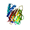
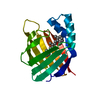
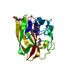
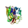
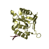
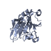
- PDB-3pve: Crystal structure of the G2 domain of Agrin from Mus Musculus -

+
Open data


ID or keywords:

Loading...

-
Basic information

Entry
Database: PDB / ID: 3pve
TitleCrystal structure of the G2 domain of Agrin from Mus Musculus
ComponentsAgrin, Agrin protein
KeywordsTRANSCRIPTION / mRNA splicing / STRUCTURAL GENOMICS / PSI-2 / PROTEIN STRUCTURE INITIATIVE / NEW YORK STRUCTURAL GENOMIX RESEARCH CONSORTIUM / NYSGXRC / New York SGX Research Center for Structural Genomics
Function / homology
Function and homology information


negative regulation of P-type sodium:potassium-exchanging transporter activity / : / negative regulation of sodium ion export across plasma membrane / A tetrasaccharide linker sequence is required for GAG synthesis / HS-GAG biosynthesis / HS-GAG degradation / positive regulation of protein geranylgeranylation / acetylcholine receptor regulator activity / regulation of axon guidance / Retinoid metabolism and transport ...negative regulation of P-type sodium:potassium-exchanging transporter activity / : / negative regulation of sodium ion export across plasma membrane / A tetrasaccharide linker sequence is required for GAG synthesis / HS-GAG biosynthesis / HS-GAG degradation / positive regulation of protein geranylgeranylation / acetylcholine receptor regulator activity / regulation of axon guidance / Retinoid metabolism and transport / ECM proteoglycans / positive regulation of synaptic assembly at neuromuscular junction / skeletal muscle acetylcholine-gated channel clustering / chondroitin sulfate binding / regulation of cardiac muscle cell membrane potential / BMP binding / sialic acid binding / plasma membrane organization / tissue development / multicellular organism development / dystroglycan binding / ATPase inhibitor activity / transforming growth factor beta binding / heparan sulfate proteoglycan binding / positive regulation of filopodium assembly / neuromuscular junction development / regulation of GTPase activity / regulation of synapse organization / receptor clustering / positive regulation of protein tyrosine kinase activity / basement membrane / regulation of cardiac muscle contraction / axonal growth cone / regulation of microtubule cytoskeleton organization / synaptic cleft / synapse assembly / positive regulation of GTPase activity / animal organ morphogenesis / regulation of protein phosphorylation / sarcolemma / Golgi lumen / positive regulation of peptidyl-tyrosine phosphorylation / positive regulation of neuron apoptotic process / positive regulation of protein binding / chemical synaptic transmission / collagen-containing extracellular matrix / transmembrane transporter binding / membrane => GO:0016020 / positive regulation of protein phosphorylation / glutamatergic synapse / synapse / calcium ion binding / cell surface / positive regulation of transcription by RNA polymerase II / extracellular space / extracellular region / plasma membrane / cytosol
Similarity search - Function
Follistatin-like, N-terminal / Follistatin-N-terminal domain-like / Factor I / membrane attack complex / factor I membrane attack complex / Domain found in sea urchin sperm protein, enterokinase, agrin / Laminin G domain / Laminin-type EGF-like (LE) domain profile. / Laminin-type EGF-like (LE) domain signature. / Laminin-type epidermal growth factor-like domai / Laminin EGF domain ...Follistatin-like, N-terminal / Follistatin-N-terminal domain-like / Factor I / membrane attack complex / factor I membrane attack complex / Domain found in sea urchin sperm protein, enterokinase, agrin / Laminin G domain / Laminin-type EGF-like (LE) domain profile. / Laminin-type EGF-like (LE) domain signature. / Laminin-type epidermal growth factor-like domai / Laminin EGF domain / SEA domain superfamily / Kazal-type serine protease inhibitor domain / Laminin-type EGF domain / Kazal-type serine protease inhibitor domain / SEA domain profile. / SEA domain / SEA domain / Laminin G domain profile. / Kazal type serine protease inhibitors / Laminin G domain / Laminin G domain / Kazal domain superfamily / Kazal domain / Kazal domain profile. / EGF-like domain / EGF-like calcium-binding domain / Calcium-binding EGF-like domain / Epidermal growth factor-like domain. / Jelly Rolls - #200 / EGF-like domain profile. / EGF-like domain signature 2. / EGF-like domain signature 1. / EGF-like domain / Concanavalin A-like lectin/glucanase domain superfamily / Jelly Rolls / Sandwich / Mainly Beta
Similarity search - Domain/homology
Biological speciesMus musculus (house mouse)
MethodX-RAY DIFFRACTION / SYNCHROTRON / SAD / Resolution: 1.4 Å
AuthorsSampathkumar, P. / Do, J. / Bain, K. / Freeman, J. / Gheyi, T. / Atwell, S. / Thompson, D.A. / Emtage, J.S. / Wasserman, S. / Sauder, J.M. ...Sampathkumar, P. / Do, J. / Bain, K. / Freeman, J. / Gheyi, T. / Atwell, S. / Thompson, D.A. / Emtage, J.S. / Wasserman, S. / Sauder, J.M. / Burley, S.K. / New York SGX Research Center for Structural Genomics (NYSGXRC)
CitationJournal: To be Published
Title: Crystal structure of the G2 domain of Agrin from Mus Musculus
Authors: Sampathkumar, P.
History
DepositionDec 7, 2010Deposition site: RCSB / Processing site: RCSB
Revision 1.0Jan 19, 2011Provider: repository / Type: Initial release
Revision 1.1Jul 13, 2011Group: Version format compliance
Revision 1.2Nov 8, 2017Group: Refinement description / Category: software / Item: _software.classification / _software.name
Revision 1.3Nov 21, 2018Group: Data collection / Structure summary / Category: audit_author / Item: _audit_author.identifier_ORCID
Revision 1.4Feb 10, 2021Group: Database references / Structure summary / Category: audit_author / struct_ref_seq_dif
Item: _audit_author.identifier_ORCID / _struct_ref_seq_dif.details
Revision 1.5Feb 21, 2024Group: Data collection / Database references / Category: chem_comp_atom / chem_comp_bond / database_2
Item: _database_2.pdbx_DOI / _database_2.pdbx_database_accession
Revision 1.6Apr 3, 2024Group: Refinement description / Category: pdbx_initial_refinement_model

-
Structure visualization

Structure viewerMolecule:
MolmilJmol/JSmol

Downloads & links

-
Assembly

Deposited unit
A: Agrin, Agrin protein
B: Agrin, Agrin protein


Theoretical massNumber of molelcules
Total (without water)40,9892
Polymers40,9892
Non-polymers00
Water4,864270
1
A: Agrin, Agrin protein


Theoretical massNumber of molelcules
Total (without water)20,4941
Polymers20,4941
Non-polymers00
Water181
TypeNameSymmetry operationNumber
identity operation1_555x,y,z1
MethodPISA
2
B: Agrin, Agrin protein


Theoretical massNumber of molelcules
Total (without water)20,4941
Polymers20,4941
Non-polymers00
Water181
TypeNameSymmetry operationNumber
identity operation1_555x,y,z1
MethodPISA
Unit cell
Length a, b, c (Å)41.620, 45.580, 51.510
Angle α, β, γ (deg.)72.280, 68.530, 65.440
Int Tables number1
Space group name H-MP1
Detailsmonomer

-
Components

#1: Protein Agrin, Agrin protein


Mass: 20494.488 Da / Num. of mol.: 2 / Fragment: G2 domain
Source method: isolated from a genetically manipulated source
Source: (gene. exp.) Mus musculus (house mouse) / Gene: Agrin, Agrin (NP_067617) / Plasmid: BS-pSGX4(BS); modified pET26b / Production host: Escherichia coli (E. coli) / Strain (production host): BL21(DE3)-Codon+RIL / References: UniProt: Q6PCM6, UniProt: A2ASQ1*PLUS
#2: Water ChemComp-HOH / water


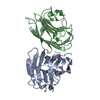
Mass: 18.015 Da / Num. of mol.: 270 / Source method: isolated from a natural source / Formula: H2O

-
Experimental details

-
Experiment

ExperimentMethod: X-RAY DIFFRACTION / Number of used crystals: 1

-
Sample preparation

CrystalDensity Matthews: 1.98 Å3/Da / Density % sol: 38.03 %
Crystal growTemperature: 294 K / Method: vapor diffusion, sitting drop / pH: 4.6
Details: 100mM sodium acetate, 15% PEG 20000, pH 4.6, Vapor diffusion, sitting drop, temperature 294K

-
Data collection

DiffractionMean temperature: 100 K
Diffraction sourceSource: SYNCHROTRON / Site: APS / Beamline: 31-ID / Wavelength: 0.97929 Å
DetectorType: RAYONIX MX225HE / Detector: CCD / Date: Feb 19, 2010
RadiationMonochromator: diamond / Protocol: SINGLE WAVELENGTH / Monochromatic (M) / Laue (L): M / Scattering type: x-ray
Radiation wavelengthWavelength: 0.97929 Å / Relative weight: 1
ReflectionResolution: 1.4→47.16 Å / Num. obs: 57584 / % possible obs: 93.3 % / Redundancy: 3.9 % / Biso Wilson estimate: 13.136 Å2 / Rsym value: 0.093 / Net I/σ(I): 7.6
Reflection shellResolution: 1.4→1.48 Å / Redundancy: 3.9 % / Mean I/σ(I) obs: 2.3 / Num. unique all: 8386 / Rsym value: 0.491 / % possible all: 92.6

-
Processing

Software
NameVersionClassificationNB
REFMAC5.5.0109refinement
PDB_EXTRACT3.1data extraction
MOSFLMdata reduction
SCALAdata scaling
SHELXCphasing
SHELXDphasing
SHELXEmodel building
CRANKphasing
RefinementMethod to determine structure: SAD
Starting model: ARP/wARP

Resolution: 1.4→36.38 Å / Cor.coef. Fo:Fc: 0.957 / Cor.coef. Fo:Fc free: 0.938 / Occupancy max: 1 / Occupancy min: 0.5 / SU B: 1.342 / SU ML: 0.053 / Cross valid method: THROUGHOUT / σ(F): 0 / ESU R Free: 0.076 / Stereochemistry target values: MAXIMUM LIKELIHOOD
Details: HYDROGENS HAVE BEEN ADDED IN THE RIDING POSITIONS U VALUES : REFINED INDIVIDUALLY
RfactorNum. reflection% reflectionSelection details
Rfree0.2289 2946 5.1 %RANDOM
Rwork0.1921 ---
obs0.194 57556 100 %-
Solvent computationIon probe radii: 0.8 Å / Shrinkage radii: 0.8 Å / VDW probe radii: 1.4 Å / Solvent model: BABINET MODEL WITH MASK
Displacement parametersBiso max: 47.33 Å2 / Biso mean: 17.1416 Å2 / Biso min: 2 Å2
Baniso -1Baniso -2Baniso -3
1-0.03 Å2-0.01 Å20 Å2
2---0.02 Å20.04 Å2
3----0.03 Å2
Refinement stepCycle: LAST / Resolution: 1.4→36.38 Å
ProteinNucleic acidLigandSolventTotal
Num. atoms2573 0 0 270 2843
Refine LS restraints
Refine-IDTypeDev idealDev ideal targetNumber
X-RAY DIFFRACTIONr_bond_refined_d0.0140.0222730
X-RAY DIFFRACTIONr_bond_other_d0.0010.021827
X-RAY DIFFRACTIONr_angle_refined_deg1.6891.9733727
X-RAY DIFFRACTIONr_angle_other_deg0.86834450
X-RAY DIFFRACTIONr_dihedral_angle_1_deg13.4585370
X-RAY DIFFRACTIONr_dihedral_angle_2_deg29.52122.566113
X-RAY DIFFRACTIONr_dihedral_angle_3_deg11.21715414
X-RAY DIFFRACTIONr_dihedral_angle_4_deg19.4981520
X-RAY DIFFRACTIONr_chiral_restr0.0890.2423
X-RAY DIFFRACTIONr_gen_planes_refined0.0120.0213122
X-RAY DIFFRACTIONr_gen_planes_other0.0010.02592
X-RAY DIFFRACTIONr_mcbond_it0.9951.51753
X-RAY DIFFRACTIONr_mcbond_other0.2641.5732
X-RAY DIFFRACTIONr_mcangle_it1.69822776
X-RAY DIFFRACTIONr_scbond_it2.413977
X-RAY DIFFRACTIONr_scangle_it3.9034.5935
LS refinement shellResolution: 1.403→1.439 Å / Total num. of bins used: 20
RfactorNum. reflection% reflection
Rfree0.346 234 -
Rwork0.322 3998 -
all-4232 -
obs--100 %

+
About Yorodumi

-
News

-
Feb 9, 2022. New format data for meta-information of EMDB entries

New format data for meta-information of EMDB entries

  • Version 3 of the EMDB header file is now the official format.
  • The previous official version 1.9 will be removed from the archive.

Related info.:EMDB header

External links:wwPDB to switch to version 3 of the EMDB data model

-
Aug 12, 2020. Covid-19 info

Covid-19 info

URL: https://pdbjlvh1.pdbj.org/emnavi/covid19.php

New page: Covid-19 featured information page in EM Navigator.

Related info.:Covid-19 info / Mar 5, 2020. Novel coronavirus structure data

+
Mar 5, 2020. Novel coronavirus structure data

Novel coronavirus structure data

Related info.:Yorodumi Speices / Aug 12, 2020. Covid-19 info

External links:COVID-19 featured content - PDBj / Molecule of the Month (242):Coronavirus Proteases

+
Jan 31, 2019. EMDB accession codes are about to change! (news from PDBe EMDB page)

EMDB accession codes are about to change! (news from PDBe EMDB page)

  • The allocation of 4 digits for EMDB accession codes will soon come to an end. Whilst these codes will remain in use, new EMDB accession codes will include an additional digit and will expand incrementally as the available range of codes is exhausted. The current 4-digit format prefixed with “EMD-” (i.e. EMD-XXXX) will advance to a 5-digit format (i.e. EMD-XXXXX), and so on. It is currently estimated that the 4-digit codes will be depleted around Spring 2019, at which point the 5-digit format will come into force.
  • The EM Navigator/Yorodumi systems omit the EMD- prefix.

Related info.:Q: What is EMD? / ID/Accession-code notation in Yorodumi/EM Navigator

External links:EMDB Accession Codes are Changing Soon! / Contact to PDBj

+
Jul 12, 2017. Major update of PDB

Major update of PDB

  • wwPDB released updated PDB data conforming to the new PDBx/mmCIF dictionary.
  • This is a major update changing the version number from 4 to 5, and with Remediation, in which all the entries are updated.
  • In this update, many items about electron microscopy experimental information are reorganized (e.g. em_software).
  • Now, EM Navigator and Yorodumi are based on the updated data.

External links:wwPDB Remediation / Enriched Model Files Conforming to OneDep Data Standards Now Available in the PDB FTP Archive

-
Yorodumi

Thousand views of thousand structures

  • Yorodumi is a browser for structure data from EMDB, PDB, SASBDB, etc.
  • This page is also the successor to EM Navigator detail page, and also detail information page/front-end page for Omokage search.
  • The word "yorodu" (or yorozu) is an old Japanese word meaning "ten thousand". "mi" (miru) is to see.

Related info.:EMDB / PDB / SASBDB / Comparison of 3 databanks / Yorodumi Search / Aug 31, 2016. New EM Navigator & Yorodumi / Yorodumi Papers / Jmol/JSmol / Function and homology information / Changes in new EM Navigator and Yorodumi

Read more