 Movie
Movie Controller

[English] 日本語
 Yorodumi
- PDB-1dkd: CRYSTAL STRUCTURE OF A GROEL (APICAL DOMAIN) AND A DODECAMERIC PE... -
+ Open data

- Basic information
| Entry | Database: PDB / ID: 1dkd | ||||||
|---|---|---|---|---|---|---|---|
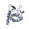
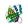
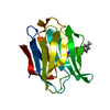
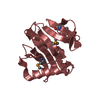
| Title | CRYSTAL STRUCTURE OF A GROEL (APICAL DOMAIN) AND A DODECAMERIC PEPTIDE COMPLEX | ||||||
 Components Components |
| ||||||
 Keywords Keywords | CHAPERONE / MOLECULAR CHAPERON / HSP60 / PROTEIN FOLDING / PEPTIDE SELECTION / PHAGE DISPLAY / PEPTIDE BINDING GROOVE FORMED BY PAIRED HELICES SUBSTRATE PEPTIDE IN BETA-SHEET | ||||||
| Function / homology |  Function and homology information Function and homology informationGroEL-GroES complex / chaperonin ATPase / virion assembly / isomerase activity / ATP-dependent protein folding chaperone / response to radiation / : / response to heat / protein refolding / protein folding ...GroEL-GroES complex / chaperonin ATPase / virion assembly / isomerase activity / ATP-dependent protein folding chaperone / response to radiation / : / response to heat / protein refolding / protein folding / magnesium ion binding / ATP hydrolysis activity / ATP binding / membrane / identical protein binding / cytosol Similarity search - Function | ||||||
| Biological species |  | ||||||
| Method |  X-RAY DIFFRACTION / SYNCHROTRON / Resolution: 2.1 Å X-RAY DIFFRACTION / SYNCHROTRON / Resolution: 2.1 Å | ||||||
 Authors Authors | Chen, L. / Sigler, P.B. | ||||||
 Citation Citation | Journal: Cell(Cambridge,Mass.) / Year: 1999 Title: The crystal structure of a GroEL/peptide complex: plasticity as a basis for substrate diversity. Authors: Chen, L. / Sigler, P.B. | ||||||
| History |
|
- Structure visualization
| Structure viewer | Molecule: MolmilJmol/JSmol |
|---|
- Downloads & links
-Download
| PDBx/mmCIF format | 1dkd.cif.gz | 126 KB | Display | PDBx/mmCIF format |
|---|---|---|---|---|
| PDB format | pdb1dkd.ent.gz | 100.3 KB | Display | PDB format |
| PDBx/mmJSON format | 1dkd.json.gz | Tree view | PDBx/mmJSON format | |
| Others |  Other downloads Other downloads |
-Validation report
| Arichive directory | https://data.pdbj.org/pub/pdb/validation_reports/dk/1dkdftp://data.pdbj.org/pub/pdb/validation_reports/dk/1dkd | HTTPS FTP |
|---|
-Related structure data


-Links
 PDBj
PDBj
- Assembly
| Deposited unit | 
| ||||||||
|---|---|---|---|---|---|---|---|---|---|
| 1 | 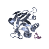
| ||||||||
| 2 | 
| ||||||||
| 3 | 
| ||||||||
| 4 | 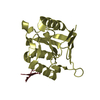
| ||||||||
| 5 | 
| ||||||||
| 6 | 
| ||||||||
| Unit cell |
|
-Components
| #1: Protein | Mass: 15729.269 Da / Num. of mol.: 4 / Fragment: APICAL DOMAIN Source method: isolated from a genetically manipulated source Source: (gene. exp.) #2: Protein/peptide | Mass: 1460.676 Da / Num. of mol.: 4 / Source method: obtained synthetically Details: the peptide was chemically synthesized and was SELECTED FROM PHAGE DISPLAY PEPTIDE LIBRARY. #3: Water | ChemComp-HOH / |  Mass: 18.015 Da / Num. of mol.: 96 / Source method: isolated from a natural source / Formula: H2O Mass: 18.015 Da / Num. of mol.: 96 / Source method: isolated from a natural source / Formula: H2OCompound details | The peptide adopts a typical beta-turn; paired alpha helices H and I, and the groove between them ...The peptide adopts a typical beta-turn; paired alpha helices H and I, and the groove between them constitute the binding site. | |
|---|
-Experimental details
-Experiment
| Experiment | Method: X-RAY DIFFRACTION / Number of used crystals: 1 |
|---|
- Sample preparation
| Crystal | Density Matthews: 2.45 Å3/Da / Density % sol: 49.81 % | ||||||||||||||||||||||||
|---|---|---|---|---|---|---|---|---|---|---|---|---|---|---|---|---|---|---|---|---|---|---|---|---|---|
| Crystal grow | Temperature: 291 K / Method: vapor diffusion, hanging drop / pH: 8 Details: PEG4K, MgCl2, TrisCl, pH 8.0, VAPOR DIFFUSION, HANGING DROP, temperature 18K | ||||||||||||||||||||||||
| Crystal grow | *PLUS Method: vapor diffusion | ||||||||||||||||||||||||
| Components of the solutions | *PLUS
|
-Data collection
| Diffraction | Mean temperature: 80 K |
|---|---|
| Diffraction source | Source: SYNCHROTRON / Site: CHESS  / Beamline: F1 / Wavelength: 0.923 / Beamline: F1 / Wavelength: 0.923 |
| Detector | Type: BRANDEIS / Detector: CCD / Date: Sep 17, 1998 |
| Radiation | Protocol: SINGLE WAVELENGTH / Monochromatic (M) / Laue (L): M / Scattering type: x-ray |
| Radiation wavelength | Wavelength: 0.923 Å / Relative weight: 1 |
| Reflection | Resolution: 2.1→25 Å / Num. all: 38529 / Num. obs: 35897 / % possible obs: 93.2 % / Observed criterion σ(I): -3 / Redundancy: 4 % / Biso Wilson estimate: 16.1 Å2 / Rmerge(I) obs: 0.077 / Net I/σ(I): 18.6 |
| Reflection shell | Resolution: 2.1→2.18 Å / Redundancy: 2 % / Rmerge(I) obs: 0.303 / Num. unique all: 3394 / % possible all: 89.9 |
| Reflection shell | *PLUS % possible obs: 89.9 % |
- Processing
| Software |
| |||||||||||||||||||||||||
|---|---|---|---|---|---|---|---|---|---|---|---|---|---|---|---|---|---|---|---|---|---|---|---|---|---|---|
| Refinement | Resolution: 2.1→25 Å / σ(F): 0 / Stereochemistry target values: Engh & Huber
| |||||||||||||||||||||||||
| Refinement step | Cycle: LAST / Resolution: 2.1→25 Å
| |||||||||||||||||||||||||
| Refine LS restraints |
| |||||||||||||||||||||||||
| Software | *PLUS Name: 'CNS' / Classification: refinement | |||||||||||||||||||||||||
| Refine LS restraints | *PLUS
|
